AI in Health care
AI in Medical Imaging: What is Coming Next

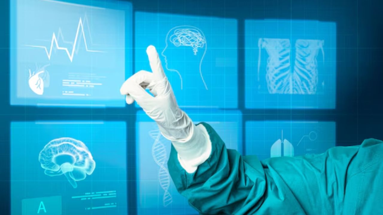
Speeds, accuracy, are vital for AI medical imaging. In case you do not know, one image can alter a diagnosis, or treatment. Artificial Intelligence will be big in healthcare, like it is in others. However, it won’t take the place of doctors, but make diagnosis and treatment faster.
AI in medical imaging is about pattern recognition at levels no human can achieve. These models analyze thousands of scans in just minutes. The result: radiology and diagnostic imaging get better clinical support.
This technology didn’t just arrive suddenly. Today, AI medical imaging products are used in hospitals, research facilities, and diagnostic laboratories globally.
Before delving deeper, let us see how AI works in medical imaging, the benefits it provides, and the future in healthcare.
What Is AI in Medical Imaging?
Artificial intelligence (AI) in medical imaging is a term that applies to a computer system that interprets the images based on the patterns it has ‘seen’ before.
These are based on machine learning and deep learning models trained on extensive datasets of labeled medical images. The system they create is more accurate after exposure to more data for the models.
Convolutional neural networks are what the majority of medical imaging AI depend on. They are very useful in image analysis, detecting edges, shapes, textures, and spatial relations. This allows AI to detect abnormalities at high precision, such as tumors, fractures, or damaged organs.
AI does not view images the way a human does. It transforms images into numbers and looks at pixel-level patterns. That’s the sort of difference that allows AI to recognize changes that are visually undetectable but important.
The output of the AI imaging system comprises classifications, probability scores, segmentation maps, and alerts. These outputs help during diagnosis, reporting, and treatment planning.
Why Medical Imaging Needs AI Support
Medical imaging demand is increasing by the day. CT scans, MRIs, X-rays, and ultrasounds are constantly imported. AI in radiology helps reduce workload by highlighting urgent cases and inaccurate research.
Human interpretations differ with experience, workload, and environmental conditions. AI offers homogeneous analysis, which is the same over time and across institutions. This uniformity is useful in the larger healthcare systems.
Technologies Behind AI in Medical Imaging
Deep learning is the basis of the majority of imaging AI solutions. These models depend on having large, interpreted datasets. The quality of data is as important as the quantity in medical image analysis.
Training datasets often contain images from different machines, patient groups, and types of clinical settings. This variation allows models to better generalize to unseen, real-world settings.
Computer vision techniques enable the AI to point out where an abnormality lies within images. For instance, segmentation algorithms delineate tumors or organs, enable physicians to quantify size, shape, and changes.
Machine learning models also integrate imaging data with clinical data. This fusion increases diagnostic confidence by providing context beyond the image alone.
Applications of AI in X-Ray Imaging
X-ray imaging is among the first domains where AI demonstrated clinical service. Chest X-rays are used routinely in diagnosing pneumonia, tuberculosis, fractures, and lung nodules.
Systems for reading AI medical imaging can interpret chest X-rays in seconds. They detect disease-associated patterns and produce probability scores, which can be used as a second opinion for radiologists in the interpretation.
Research shows that AI improves detection rates for e.g., lung nodules and pneumothorax. In emergency medicine, this can make a big difference in patient outcomes. AI is now bolstering screening programs in regions where radiology expertise is scarce. This use case expands access to diagnostic care for populations in need.
AI in CT Scan Analysis
CT scanning generates a massive amount of data. A CT scan may include hundreds of slices, and meticulously reviewing each one is a time-consuming process. These sequences are assessed in parallel by AI CT scan applications. They recognized anomalies, such as internal bleeding, pulmonary embolism, and tumors.
AI is essential in stroke care. Automated systems identify large vessel occlusions and notify the clinical team instantly. This pace decreases treatment latencies and enhances survival rates.
Also in CT imaging, AI is being explored for quantitative analysis to more accurately track the progression of disease and response to treatment.
AI in MRI Reading
MRI offers excellent soft tissue imaging, but takes more time to acquire and interpret. AI streamlines the two. Reconstruction capabilities of AI MRI solutions to address this, shortening the scan time while preserving image quality. Faster scans enhance patient comfort and throughput.
When it comes to interpretation, AI detects abnormalities in the brain, spine, joints, and organs. It aids the diagnosis of diseases such as multiple sclerosis, brain tumors, and ligament injuries. These tools increase precision and reduce reader variability, notably in challenging cases.
AI In Ultrasound Imaging
Operator dependence is strong in ultrasound imaging. The quality of images or interpretation can differ quite a bit from user to user. AI-controlled ultrasound devices to assist in image acquisition. They instruct users to hold probes at the right angle(s) to obtain diagnostic images.
Moreover, AI enables expanded ultrasound applications in primary care and in field locations, where specialist availability is limited.
The AI Revolution in Radiology
The application of AI in radiology is not confined to image reading. It is a resource for imaging job flows during the lifecycle. AI software triages incoming cases and prioritizes critical findings. That raises this urgent case priority immediately, which is great.
Measurements are extracted, and structured reports are populated using automated reporting solutions. This standardization streamlines the reporting process and saves time.
Quality control: AI is also used to identify incomplete studies and technical errors before they are interpreted.
Clinical Evidence for AI Medical Imaging
- Clinical validation is Needed. Several AI imaging products are tested extensively in retrospective and prospective studies.
- Peer-reviewed studies indicate that, for some diseases, AI used in addition to radiologists increases sensitivity and specificity. Combined performance is guaranteed to be superior to individual performances.
- The FDA and CE mark regulatory approvals require the submission of proof of safety and efficacy. These approvals signify clinical, not experimental, application.
- Continuous post-market surveillance assesses real-life performance and limitations. AI Is Decision Support, Not Replacement
The fundamental tenet of AI in medical imaging is augmentation, not replacement. AI doesn’t make the final clinical decisions.
Radiologists review AI outputs within a clinical context. They have patient history, symptoms, and other test results. This human oversight prevents blind faith in algorithms and ensures accountability. AI is a tool, not an expert. This shared model is consistent with professional ethics and preserves patient trust.
Bias and Data Quality Issues
The AI systems are trained with historical data. If the data used to train the model is biased, the model might also be biased.
Bias may also be introduced in the medical imaging process through population or modality-biased sampling. This constraint limits the applicability of the results.
Software vendors are tackling this by making their datasets more diverse and by performing external validation studies. Being transparent about the limitations of a model is paramount for safe use in clinical practice.
Data Privacy and Security Measures
Medical images are associated with sensitive patient information. They also must adhere to AI systems’ data protection requirements. Data security, encryption, and de-identification need to be implemented. Healthcare providers need to be considered.
However, additional protections for cloud-based AI systems against unauthorized access are also necessary. Trust is based on good governance and observance of ethical standards.
AI-Based Imaging Products
AI medical imaging is subject to developing regulations. Authorities now require an unambiguous demonstration of performance, safety, and clinical benefit.
Adaptive algorithms are a new regulatory frontier. Models that evolve over time need to be continuously monitored. These actions safeguard patients and, at the same time, are supportive of responsible innovation.
Explainability in AI Medical Imaging
- Health care providers need to know the reasons an AI system makes a particular recommendation. Explainability adds to the trust in and adoption of the system.
- Visualization tools reveal which regions of an image are contributing to the predictions of an AI. These maps allow radiologists to confirm detections.
- Explainable AI contributes to education and quality control.
- It’s difficult to apply AI in the clinic, because you have no explainability for it.
AI and Multimodal Imaging Analysis
Contemporary diagnosis frequently uses several imaging techniques. AI processes data from CT, MRI, and PET scans. Multimodal analysis enhances the accuracy of diagnosis by integrating complementary information.
AI also connects imaging findings to pathology, genomics , and clinical. This fusion enables precision medicine and individualized therapy planning.
Effect on Patient Outcomes
The primary objective for AI in medical imaging is better patient care. Earlier diagnosis means earlier treatment. Greater accuracy leads to unnecessary procedures.
AI enables standardized care within organizations and at different centers, reducing disparities. In addition, AI contributes to a more efficient healthcare systems.
Barriers to Training and Adoption
Training to use AI is a part of effective adoption. Radiologists must be able to trust and understand the systems. There is need for constant learning.
Economic Effects of AI in Imaging
The AI in medical imaging can lead to cost savings by increasing speed and quality, and decreasing the number of repeated scans. As a result, early detection of diseases will save on the related costs of treatment. But costs for initial investment and integration must be factored in.
AI in International and Remote Medicine
In countries with few trained radiologists, AI is enhancing diagnostic accessibility. Cloud-based imaging AI facilitates remote reading and screening programs. This functionality enhances rural healthcare and global health initiatives.
AI doesn’t replace specialists; it extends their reach.
Prospects of AI in Medical Imaging
- AI will also proceed towards online, adaptive systems.
- Integration with clinical decision support will intensify.
- Regulation, ethics , and explainability will influence the development priorities.
- Emphasis will still be on performing safe evidence-based improvements in patient care.
What is coming Next?
AI in Medical Imaging Research
Research is what pushes AI in medical imaging forward. Testing in academic centers and teaching hospitals largely occurs for new algorithms. These settings enable comparison of AI performance to expert radiologists on validated datasets. So, it is vital that changes are measured and not just assumed.
Clinical trials are incorporating AI applications in imaging workflows. Researchers evaluate the impact of AI on diagnostic performance, reading time, and medical results. Information from these investigations guides regulation and clinical use.
AI medical imaging tools cannot be trusted long-term without robust research underpinning them. Open data also enables faster innovation.
AI and Rare Disease Detection
AI-based medical imaging platforms can detect unusual patterns that even doctors may not see. Using aggregated global datasets to train, AI can pick up on subtle imaging signs associated with rare diseases.
Radiographers receive AI alerts on possible alternative diagnoses. These pushes are for further work and not for final answers. This adjunctive function enhances diagnostic confidence without overriding the clinician Judgment.
Quality Assurance and Error Reduction
Medical imaging diagnostic errors may be caused by fatigue, time constraints or subtle abnormalities. Quality assurance in radiology is influenced by availability of AI as a standardized experience second reader.
AI systems mark inconsistencies, unnoticed findings, or suspicious trends. Radiologists may also examine these warnings prior to completing reports.
Support tools for quality assurance, high quality, and facilitate peer review and education. The institution employs this AI feedback to detect trends in misinterpretations. In the long run, this translates into safer and more dependable diagnostic services.
AI for Pediatric Medical Imaging
Pediatric imaging protocols are optimized using AI. In X-ray and CT scans, AI enables dose reduction without compromising diagnostic quality. This is important because the long-term risk can be reduced.
Pediatric-trained AI models enable accurate interpretations for patients of all ages. This expertise increased confidence for diagnoses that involved growth-related changes. Moreover, pediatric radiologists have access to solutions designed for their specific clinical needs.
The Ethical Responsibility in AI Imaging
Ethical issues influence the development and use of AI in medical imaging. Transparency, accountability, and patient safety remain pressing issues. Physicians need to recognize the boundaries of the system and not place blind trust in what the algorithms report.
Clear documentation and performance metrics enable responsible use. Patients are entitled to that, too. When AI assists with diagnosis, institutions can be transparent about that role.
Ethical AI plays a key role in building trust by ensuring technology works with professional standards and patient expectations.
Continuously Learning and Updating Models
The world of medicine keeps changing, and so should the AI systems. Continuous learning enables imaging AI to improve. Model updates must be validated to verify that performance does not degrade. Healthcare Providers need to monitor version changes and re-assess outcomes.
And regulators are increasingly demanding post-deployment monitoring. This supervision ensures that AI is safe and effective throughout its existence. An update responsibly manages to strike the right balance of keeping innovation fresh and stable in a clinical setting.
Integration with HIS
AI medical imaging solutions should be fully compatible with established systems. PACS and EHR are vital to the imaging workflow. The standards ensure AI solutions are enabled to work across different vendors and organizations. Seamless integration enables adoption and impact in the real world.
Training the Next Generation of Radiologists

People are taught to question AI outputs rather than accept them uncritically. This expertise allows you to maximize your commitment to diagnosis. Exposure during training enhances acceptance of new technology and minimizes barriers to adoption in professional life.
Future radiologists will routinely use AI as a requirement in imaging care.
Defining Success Beyond Accuracy
- Measuring success by accuracy alone is insufficient for AI in medical imaging. Clinical value goes beyond efficiency and usability, and also includes patient outcomes.
- Hospitals are checking if AI can speed up image reporting, make work smoother, and help with quicker treatment.
- User experience matters. Any products that interfere with workflow will have difficulty gaining traction, no matter how accurate they are.
Conclusion
The evidence, regulation, and clinical experience continue to advance. The best systems are those that support clinicians without displacing human effort. No doubt, Artificial intelligence increases speed and consistency.
AI in medical imaging is no longer investigative. It is a rational solution with quantifiable clinical benefits as long as it is used correctly. Very importantly, AI enhances, rather than replaces, clinical findings.

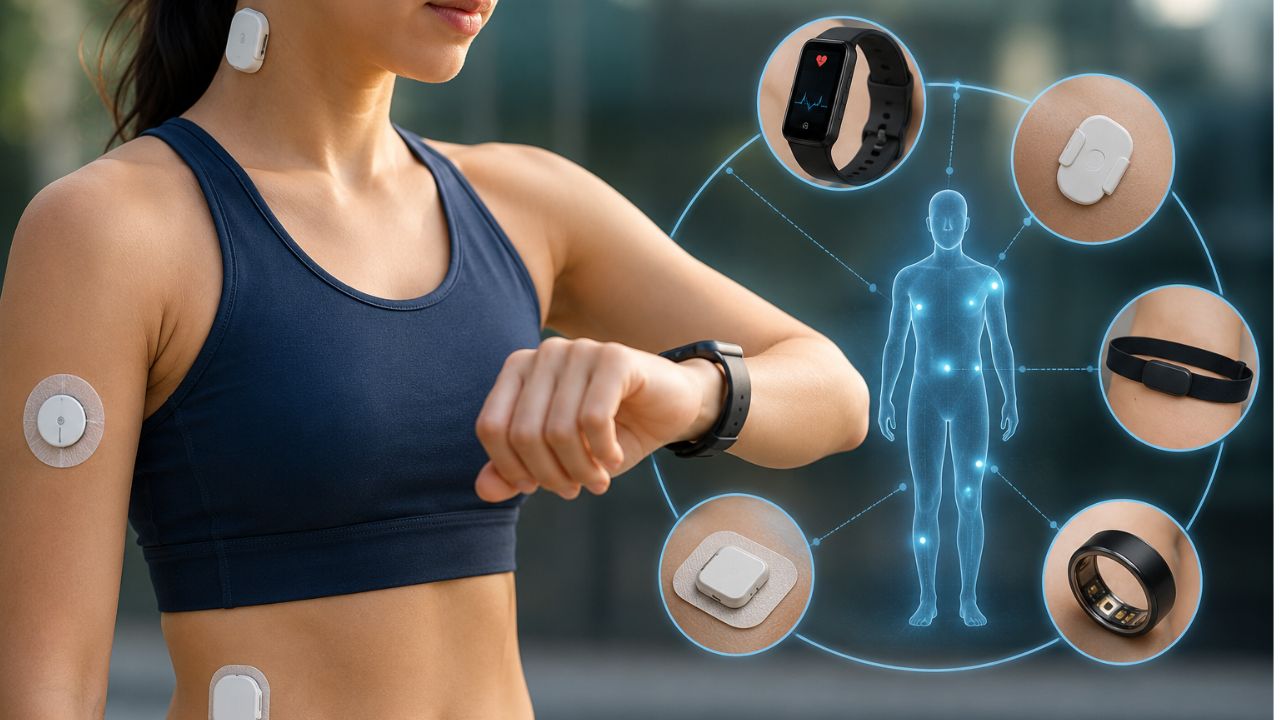
The healthcare sector is undergoing a huge transformation. Now, many are shifting from reactive medicine to preventative care. The technology spearheading this change are wearable biosensors. These are not just fancy devices; they are advanced medical devices you can wear on your wrist or stick to your skin like a band-aid.
In this article, we’ll take a look at how the sensors function, their categories, and the top wearable devices available.
What is a Wearable Biosensor?
Wearable biosensors are small electronic devices that detect biological or physical signals from the body and transmit the information to a smartphone, smartwatch, tablet, or medical device.
The simple description of this device is that it “reads” your body. It takes biological signals such as your sweat chemical composition or your heart rate, converts them into digital information, and you can access it on your smartphone or share it with your doctor.
The beauty of a wearable biosensor is that it can be continuously monitored. In the past, if you wanted to check your blood sugar or heartbeat, you had to visit a clinic. However, in recent times, these biosensors can capture your health even while you eat, sleep, work, and exercise.
How Does Wearable Biosensors Work?
- Biological Fluids: It checks liquids like sweat, saliva, and interstitial fluid (the fluid between cells).
- Biomarkers: It looks for specific markers like glucose for diabetes or lactate for muscle fatigue.
- Physiological Conditions: It monitors heart rate, oxygen, and breathing.
Why Healthcare Monitoring Is Becoming More Important
We are no longer waiting for things to go wrong before we act, and this is why monitoring is a vital part of modern medicine. When we monitor data in our bodies, we can detect disease early, prevent chronic disease, and live longer.
Here is why we should monitor our health.
The Rise of Chronic Conditions
Globally, there is an increase in chronic lifestyle diseases such as diabetes, hypertension, and heart disease. These conditions don’t appear all of a sudden. They develop over years with or without symptoms that we miss during routine yearly check-ups. However, wearable biosensors can detect these conditions.
Someone at risk of diabetes can notice how their biological fluids respond when they eat sugary meals. This real-time data helps one to modify what they consume.
Moving from Reactive to Proactive Care
The old medical model was reactive. Once you feel pain, you go for a lab test and then commence treatment. At present, we focus on proactive care. When a patient uses a wearable biosensor, they get quick results and also optimize their health.
If you track biomarkers like your heart rate or sleep quality, it will show you how stress and diet affect your physiological conditions.
Reduces the Burden on Hospitals
Constant healthcare monitoring allows doctors to keep track of high-risk patients while they are at home. If a heart patient’s patch detects an irregular rhythm, the hospital can be alerted immediately. This can prevent a minor issue from turning into a major emergency.
Precision Medicine and Personalization
Every human body is different. A diet that works for one person might cause inflammation in another. Through the use of a specialized wearable biosensor, we can move toward precision medicine.
This means treatments and lifestyle advice works for a specific data. Whether it is a device that tracks your oxygen levels or a sensor that monitors your hydration via sweat, the treatment plan is for a particular individual..
The Power of Continuous Data
When a doctor asks, how have you been feeling for the last three months? Most of us can only give a vague answer. But, a wearable biosensor provides an objective, 24/7 record of your body’s performance like:
- Real-time Alerts: It can alert you to move if your circulation is poor.
- Sleep Insights: It can identify signs of sleep apnea as it tracks blood oxygen.
- Stress Management: It can detect spikes in cortisol or skin conductance, telling you when it is time to take a breath.
Main Components of Wearable Biosensors
This comprises several parts that make the device work well. The components are:
Biological Recognition Element
This layer identifies the target substance or biological activity.
Examples:
- Enzymes
- Antibodies
- DNA fragments
- Living cells
Sensor
The sensor detects chemical, electrical, or physical changes
Signal Processor
The processor converts raw signals into digital information.
Communication Module
Bluetooth, Wi-Fi, or cellular technology sends data to connected devices.
Software
The application shows results, stores records, and provides alerts when necessary.
Categories of Wearable Biosensors
Biochemical Wearable Biosensors
These devices search for chemical changes in the body. They provide continuous information throughout the day.
Interstitial Fluid Sensors
Interstitial fluid sits under the skin and contains valuable health information.
Continuous glucose monitors (CGMs) use a small flexible filament placed just under the skin. The sensor measures glucose levels every few minutes.
Examples:
- Abbott FreeStyle Libre
- Dexcom G6
These devices help people with diabetes maintain stable glucose levels without frequent finger-prick testing.
Benefits:
- Continuous glucose tracking
- Proper diabetes management
- Few blood tests
Sweat Analysis Sensors
Electronic skin patches and wearable tattoos measure important biomarkers such as the following:
- Lactate
- Glucose
- Sodium
- Potassium
- Chloride
Uses:
- Monitors Hydration
- Sports performance
- Exercise recovery
- Prevent heat stress
Tear and Saliva Biosensors
These systems can monitor:
- Tear glucose
- Intraocular pressure
- Stress hormones
- Infection markers.
Electromechanical & Biophysical Sensors
This type of wearable biosensor tracks movement and electrical signals. Usually, they are thin, stretchable, and feel like a second skin.
ECG and Heart Rate Sensors
These sensors record electrical activity from the heart.
Many smartwatches and medical patches can monitor:
- Heart rate
- Heart rhythm
- Blood oxygen (SpO₂)
- Skin temperature
Examples:
- Apple Watch
- Medical ECG patches
- Portable cardiac monitors
Respiration and Movement Sensors
Wearable chest patches and smart clothing measure body movement.
They can monitor:
- Breathing rate
- Walking patterns
- Posture
- Sleep movement
- Activity levels
Technologies behind Modern Wearable Biosensors
Although the technologies in the tools are still improving. Here is what they contain:
Flexible Materials
Manufacturers now use lightweight materials that move naturally with the body.
Examples include:
- Biocompatible polymers
- Hydrogels
- Flexible electronics
- Electronic textiles
Edge Artificial Intelligence
Many modern sensors include built-in artificial intelligence.
Instead of sending every reading to cloud servers, the device processes information immediately.
Edge AI helps to
- Remove motion interference
- Improve signal quality
- Detect abnormal readings
- Deliver faster alerts
- Reduce battery use
What Biomarkers Can Wearable Biosensors Measure?
Each sensor detect different health symptoms.
The known biomarkers are:
- Glucose
- Lactate
- Cortisol
- Electrolytes
- Oxygen saturation
- Body temperature
- Heart rhythm
- Respiratory rate
What Are Biological Fluids?
These fluids carry essential health information throughout the body.
Examples:
- Blood
- Interstitial fluid
- Sweat
- Tears
- Saliva
- Urine.
The Physiological Conditions that Wearable Biosensors Monitor
These devices monitor several physiological conditions, such as:
- Heart rate
- Blood oxygen
- Body temperature
- Sleep quality
- Physical activity
- Breathing rate
- Blood glucose
- Stress response
- Hydration
- Blood pressure trends
Top 5 Wearable Medical Devices
To help you understand the current market, here are the five essential wearable medical devices.
Continuous Glucose Monitors (CGMs)
A CGM is a sophisticated wearable biosensor for people living with diabetes. It doesn’t use the traditional finger-prick blood tests.
Rather, a small patch with a tiny, flexible filament that sits just under the skin. In addition, it measures glucose levels in your biological fluids.
Real-Time Data: It sends sugar levels directly to a smartphone or insulin pump.
- Trend Tracking: It shows if your levels are rising or falling after a meal or exercise.
- Safety: It alerts the user (and often their doctor) if sugar levels become too high or low.
ECG Monitoring Patches
While some watches can take a heart reading, a dedicated ECG (electrocardiogram) patch is recommended. These are small, paste strips you stick directly to your chest.
They are much more accurate than wrist-worn devices because they are closer to the heart and are not affected by arm movement.
- Heart Rhythm Detection: They are used to find irregular heartbeats, such as atrial fibrillation (AFib).
- Extended Use: A patient can wear a single patch for up to 14 days. It captures heart data during sleep, work, and exercise.
- Wireless Connectivity: Modern patches stream data to a cloud-based platform where cardiologists can review the findings in real time.
Smartwatches with wearable biosensors
Advanced versions from brands like Apple, Samsung, and Google have medical-grade sensors cleared by health authorities. These devices monitor several biomarkers through light-based technology (PPG) and electrical sensors.
- Blood Oxygen (SpO₂): They measure the oxygen your red blood cells carry.
- Sleep Vitals: They can detect disruptions in breathing that may suggest sleep apnea.
- Vital Alerts: They can notify you of an unusually high or low resting heart rate.
Wearable Blood Pressure Monitors
There are wearable blood pressure monitors that fit on your wrist like a watch. These devices use advanced “oscillometric” technology to inflate a small cuff inside the strap.
- Hypertension Management: They allow multiple readings which helps doctors understand “white coat hypertension” (stress caused by being in a clinic).
- Automatic Logging: Every reading is saved in an app. It makes it easy to see how medication and diet affect your blood pressure.
Wearable Cardiac Rhythm Monitors
Some of these take the form of incredibly thin, stretchable smart stickers or even smart clothing. They depend on the electrical signals of the heart over weeks or months to catch rare but dangerous cardiac events.
- Post-Surgery Care: These are used for patients who are just recovering from major heart procedures.
- Biophysical Tracking: They monitor heart rate variability (HRV).
- Discreet Design: Most of these devices are so thin that they can be worn under regular clothing without being noticed.
What Are the Examples of Wearable Biosensors?
There are plenty of them. The popular ones are:
- Abbott FreeStyle Libre
- Dexcom G6
- Apple Watch
- LifeSignals wearable patch
- Smart ECG patches
- Smart rings
- Electronic skin patches
- Smart contact lenses under development
- Smart health bands
Benefits of Wearable Biosensors
A modern wearable biosensor uses materials like hydrogels and e-textiles to stay comfortable. They also use Edge AI. This means the device cleans the data on your arm before sending it to the cloud.
The Key benefits are:
- Continuous healthcare monitoring
- Detects disease on time
- Better chronic disease management
- Promotes personalized treatment
- Improves patient engagement
- Support remote healthcare support
- Better medication monitoring
- Real-time alerts
- Greater convenience
Where to use wearable biosensors
The devices can be used in managing and monitoring the following medical conditions:
Diabetes Management
Continuous glucose monitors help patients maintain their glucose levels.
Benefits:
- Better insulin adjustment
- Improved glucose control
- Fewer emergency cases
Cardiovascular Care
Healthcare providers can review patient data remotely and respond almost immediately.
Uses are to:
- Detect Arrhythmia
- Monitor heart failure.
- Track blood oxygen.
Hypertension Monitoring
Wearable devices help patients follow blood pressure trends over time. However, many still require cuff calibration.
Chronic Kidney Disease
Researchers now combine biochemical monitoring with artificial intelligence to identify changes that may point to kidney problems.
This may support earlier intervention and better disease management.
Athletic Performance
Professional athletes and fitness enthusiasts use wearable devices every day.
They track:
- Lactate levels
- Hydration
- Recovery
- Muscle fatigue
- Heart rate
- Training intensity.
Preventive Healthcare
The early signs may include:
- Irregular heartbeat
- Low oxygen levels
- Fever
- Reduced activity
- Abnormal sleep patterns
This new era of healthcare is personal, data-driven, and proactive.
Challenges of Wearable Biosensors
Although the technology provides real-time health data, there are a few concerns, such as
- Data privacy concerns
- Sensor calibration
- Motion interference
- Causes skin irritation
- Wireless connectivity issues
- Regulatory approval
Read also: Cloud Computing in Healthcare: Applications and Benefits
Final Thoughts on Wearable Biosensors
We are moving away from the days of guessing why we feel tired or waiting for a yearly check-up to uncover a problem. These small patches and smart devices act like a quiet partner, watching over our vitals while we live our lives.
Whether you want to manage a condition like diabetes or are just trying to sleep better, having this data at your fingertips is empowering. It makes you be in charge of your health. This new era of healthcare is personal, data-driven, and proactive.
Perhaps we should start with this. Which chronic disease are you having? This will help us suggest a perfect wearable biosensor for you.
I hope this was helpful. For more updates, kindly follow us on our social media handles.
While many of us can relate, living with knee pain is not something you should take lightly. If you are living with osteoarthritis, you know what it feels like when your joints start grinding like old engine parts. Simple movements like walking or getting out of a chair become a struggle every single day. Chronic knee pain steals your joy and makes you feel much older than you are. But today, robotic knee replacement surgery can help. With this new technology, surgeons can now fix your knees and get you back to normal mobility.
Chronic knee pain caused by the progressive wear and tear of osteoarthritis is more than a physical illness. As cartilage in the knee joint breaks down, it creates bone-on-bone friction. When conservative treatments like physical therapy and injections do not provide relief, the thought of traditional surgery can seem discouraging.
However, things have changed in the world of orthopedics. Today, we are experiencing robotic-assisted knee replacement, a new type of knee replacement that allows doctors to bypass the limitations of traditional surgery.
What is Robotic Knee Replacement?
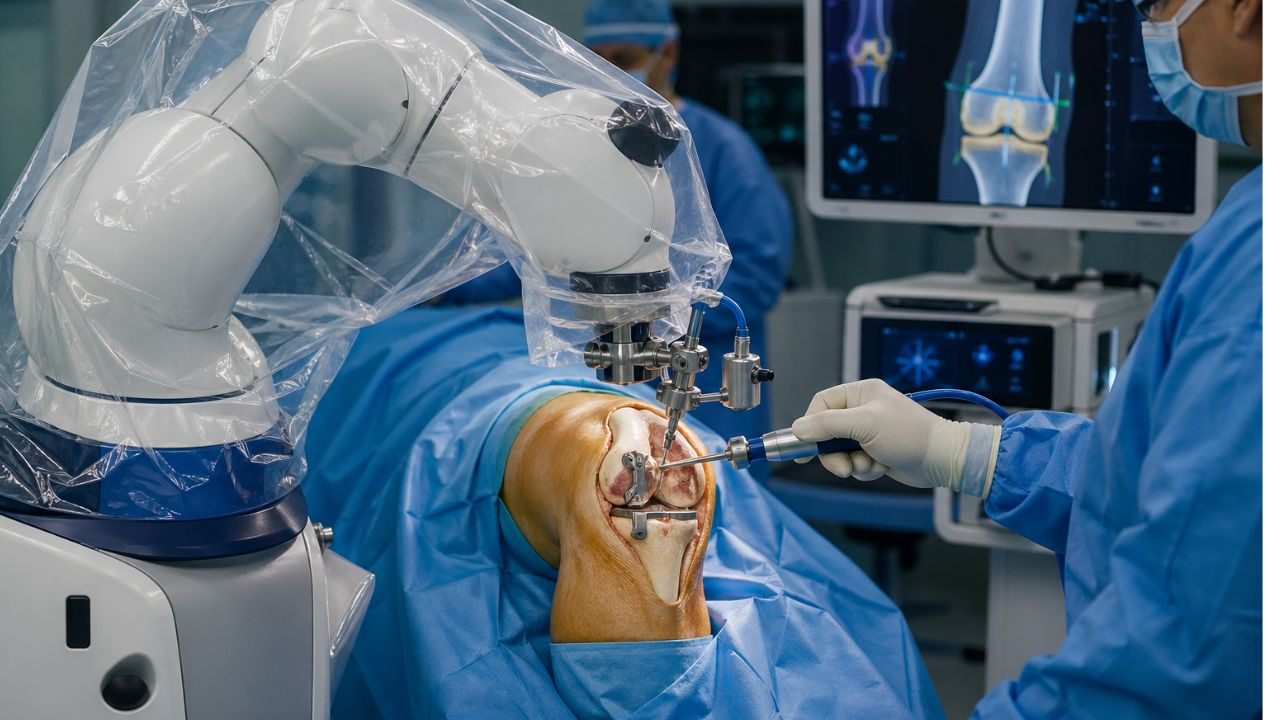
Before even stepping into the operating room, the surgeons take a CT scan of your knee and create a 3D digital map of your bone structure. Because every patient’s anatomy is slightly different, this map allows the surgeon to design the exact size and placement of your new joint. They see every single detail about your knee.
The robotic arm is used by the surgeon to remove the bone and cartilage damaged during the surgery. The robot follows a digital plan that was generated from your scan. If the surgeon moves the tool even a millimeter outside the planned area, it creates resistance or stops completely.
This prevents the surgeon from damaging the healthy tissue or removing too much bone. It is like a high-tech GPS for your knee to keep it on the right track.
It ensures that your new joint fits better. In a traditional knee replacement, the surgeon uses a manual tool and their own sight to fix the new joint. Experienced surgeons can do this very well, but the robotic system makes it more accurate.
Robotic Knee Replacement vs. Manual: Which Knee Surgery is Better?
When you have persistent knee pain, the next thing that comes to your mind is to go for surgery. In this situation, you have two options.
Manual surgery or robotic-assisted surgery
Both approaches are safe. However, how the surgeon gets there is different.
Manual Knee Replacement: The Traditional Standard
During this surgery, the surgeon uses handheld instruments to position the new joint.
They use physical guides to make the necessary cuts in the bone. The success or failure of this operation will also depend on the surgeon’s experience.
Robotic Knee Replacement: The High-Tech Assist
Robotic surgery does not mean a machine does the operation and the doctor is just watching. The surgeon is still in charge. The robot’s just a tool to help with accuracy.
A computer makes a 3D map of your knee. The robotic arm directs the surgeon to stay within the lines of that map during the operation. This prevents the surgical tool from going out of the planned area. This allows the surgeon to position the new joint with extreme accuracy, sometimes to the millimeter.
The Similarities
1. Accuracy and Fit
The robot provides a level of accuracy that the human eye may not achieve. Because all knees are unique, the robot allows for a personalized approach to knee replacement surgery based on your individual anatomy. This often leads to a more “natural” feeling knee once it heals.
2. Recovery Time
There is a somewhat speedier recovery, if only a slight one, for those who have robotic surgery. The cuts are more accurate, so there is less trauma to nearby tissue and ligaments. This results in less swelling and pain in the days after surgery.
3. Long-term end points
Both surgeries have good long-term success rates. Most patients are happy with their results, no matter what method is used.
Is Robotic Surgery Always Better?
Not necessarily. The most important thing is not the machine, but the person operating it. A skilled surgeon who has performed thousands of manual replacements will probably give you an excellent result. In addition, robotic surgery costs more. Not every hospital has this technology, and insurance coverage can sometimes be different.
Which One Is Right for You?
If you have a complex deformity in your knee, a robot’s precision might be a big help. For a common example of wear-and-tear, manual surgery is a proven option.
Here’s what you should do next:
- See if your insurance covers the robotic-assisted version.
- Inquire about the specific recovery time for your health level.
Do Surgeons put Patient to Sleep for Robotic Knee Replacement?
As you prepare for a robotic knee replacement, know this. You’ll not experience any sensation during the operation. Many people believe that general anesthesia is necessary. There’re options based on your health and your surgeon’s recommendations.
The first option is general anesthesia, a traditional method where you will remain unconscious throughout the surgery. The other is a spinal block with “twilight sleep.” The medical team administers anesthesia from the waist down to prevent pain. They also give sedative medication through an IV, which induces a deep sleep. While this is on, you remain unaware of the sounds and movements in the operating room.
The presence of the robotic system does not alter the anesthesia process. It serves as a precise tool that assists the surgeon in mapping your bone structure and positioning your new joint. Regardless of being fully asleep or in a sedative-induced state, the robotic system works under the surgeon’s command.
Ideally, you should consult an anesthesiologist who will assess your cardiovascular and pulmonary health, to determine the safest anesthesia option.
Who can have a Robotic Knee Replacement?
Surgeons often advise robotic assistance for individuals with:
Severe Osteoarthritis: The leading cause for surgery, where knee cartilage deteriorates. This results in stiffness and acute pain. Robotic systems enable surgeons to map your bone structure and position the implant correctly.
Complex Knee Anatomy: Bone shapes are different among individuals. The robot generates a 3D model of your knee before any incisions.
Persistent Pain and Stiffness: Constant morning stiffness or a dull ache even at rest may indicate irreparable joint damage. If such pain affects your sleep, a knee replacement might be needed.
General Health: Surgeons assess if patients are fit for anesthesia and ready for the recovery process. You don’t need to be an athlete, but you should have sufficient strength to move soon after the procedure.
Robotic Knee Replacement: What Happens After Surgery
How well your new knee functions in your day-to-day activities depends on your recovery efforts.
The Initial 48 Hours
Once you wake after the surgery, a physical therapist will likely assist you to stand and walk a few steps with a walker.
During these days, the focus is on managing pain and maintaining proper blood circulation. You must adhere to a pain medication schedule to prevent discomfort, and nurses will show you simple ankle exercises you can do in bed. These exercises help reduce swelling and prevent blood clots.
Rehabilitation
While the robot ensures perfect alignment of the joint, your muscles needs to adjust. Physical therapy commences after the surgery. You’ll be tasked with exercises to fully extend your leg and bend it to a certain degree. Regular movement helps to avoid scar tissue that can restrict joint mobility. Neglecting these exercises may lead to stiffness.
Long-Term Prospects
Information on robotic knee replacements show high rates of patient satisfaction. The robot provides a “custom” fit based on your bone structure, resulting in many patients feeling their knee is more natural than expected.
Most individuals resume low-impact activities, such as walking, cycling, or swimming, within six to twelve weeks. These knee replacements shows that most remain functional for at least 20 years.
Read also: Best Surgical Lights: LED Operating Room Lights
Conclusion
Robotic knee surgery provides your surgeon with a state-of-the-art assistant for enhanced accuracy. Before the operation, a scanner generates a 3D model of your leg. This blueprint enables the surgeon to strategize each incision in accordance with your unique bone structure.
During the surgery, the robotic arm keeps the instruments aligned, thereby ensuring a perfect fit for the new joint. This care prevents the surgeon from deviating even slightly from the plan. Such meticulous detail safeguards your healthy ligaments and contributes to more natural movement.

In the last few years, there have been exciting developments in medical imaging. Film X-ray was the traditional way, but recently, some health facilities are using digital X-ray for their radiography. Digital X-ray vs film X-ray: Which one do you consider the best?
This question is quite technical, so I don’t expect a straight answer. Many industries are embracing technology to boost efficiency the health sector included. But how do these two imaging technologies work? Film X-rays produce consistent, reliable diagnostic images; on the other hand, the digital X-ray system has higher speed, quality images, and easy storage and management.
But it turns out technology has made medical imaging work differently. Instead of exposing film, a digital X-ray system uses a sensor to capture the radiation, which is then converted directly into electronic images. The device transmits the information to a computer, and the result is displayed on the screen a few seconds later.
But is digital always the better choice? And what is it exactly that differentiates it from the traditioner imaging system?
What is a digital X-ray?
Digital X-ray is a form of imaging where radiographic images are captured electronically rather than relying on traditional black and white photographic film.
As an alternative to exposing X-ray film, a sensor or digital detector is used to capture the radiation signals. The signals are then converted by the detector into digital data. A computer processes the data, and the images are available on a screen in a matter of seconds. It omits some steps that are traditionally found in radiography.
In film imaging, the technicians have to develop the film to see the outcome. Digital imaging excludes that step. A typical digital X-ray system includes the following:
- An X-ray machine
- A digital sensor or detector
- Computer software for image processing
- A workstation for viewing and storing images
When an image is taken, it displays on the computer monitor immediately. With this, radiographers can adjust the brightness, enlarge the details, and then send the files. This easy workflow is one of the reasons healthcare providers prefer digital radiography.
How Traditional Film X-Ray Works
Film radiography is the orthodox method radiologists used before the introduction of digital x-ray. Although some clinics still use them . In this system, the machine sends X-ray radiation through the body. The radiation reaches a sheet of film placed behind the patient.
Different tissues absorb radiation at different levels. Bones absorb more radiation, while soft tissues allow more rays to pass through. These variations create patterns on the exposed film.
The exposed X-ray film must go through chemical development in a darkroom. The process involves developer chemicals, fixer solutions, and washing stages.
After processing, the film dries, and then it shows the final image. To be honest, this workflow takes several minutes. In busy clinics, it can slow down patient flow.
Still, film radiography has one key advantage. It produces extremely high-resolution images when handled correctly. That dependability is why some institutions continue to use film systems.
The Difference Between Digital X-Ray vs Film X-Ray
The only thing that separates digital and film imaging is the way images are captured, processed, and stored. Let’s make this simple.
Image capture technology
Film X-ray uses photographic film to capture an image of radiation exposure.
Digital radiography, however, captures the radiation signal with an electronic sensor or detector. This is the basic level-out distinction between the two systems.
Image Processing
Film images must be developed with a chemical before the images can be captured. But, a digital X-ray system processes images immediately with a computer software. It only takes a few seconds for a technician to look at the images. There is no wait period.
Storage and Access
The storage is another salient point. Film images are stored or accessed manually from their storage rooms. Digital images are stored in hospital databases or on cloud storage. It makes retrieval much faster and easier.
Image Sharing
Film images must be physically transported or scanned. Digital images can be shared instantly with other specialists through electronic systems.
Workflow Efficiency
Film imaging involves multiple manual steps.
Digital imagine has a straightforward workflow.
This difference improves productivity in high-volume clinics.
Digital X-Ray Machine vs. Film X-Ray Machine
Both technologies focus on similar core equipment. The machine that produces X-rays are somehow identical. However, the imaging components are not the same.
Film X-Ray Machine Setup
A traditional film system typically includes:
- X-ray generator
- Film cassette
- Intensifying screen
- Film processing equipment
- Chemical developer and fixer solutions
Technicians must handle film carefully to avoid exposure errors.
Digital X-Ray Machine Setup
A digital X-ray system replaces film cassettes with detectors.
These detectors may include:
- Flat panel detectors
- CCD sensors
- CMOS sensors
- The sensor captures radiation data and sends it directly to a computer.
- The system then converts the data into diagnostic images almost instantly.
- This simplified process is one reason digital radiography continues to expand worldwide.
X-Ray Film Digitizers: How Conversion Works
Several facilities want to upgrade but already have large archives of film images. This raises an important question: can X-ray film be digitized for modern systems?
The answer is yes.
Clinics usually convert old film records into digital files through scanning technology. Specialized scanners capture the film image and convert it into digital data. Once scanned, the images can be stored in electronic databases.
This process allows facilities to keep historical records without maintaining large film storage rooms.
Digitizing film archives also help physicians have access to files especially if there is need to compare recent and old image.
Advantages of Digital X-Ray
There are so many benefits of digital radiography. They include the following:
Faster Image Results
Digital systems display images within seconds. Technicians can confirm image quality immediately and repeat scans if necessary.
This reduces patient waiting time.
Lower Radiation Exposure
Many digital systems require less radiation than traditional film imaging. Advanced sensors capture more data from smaller radiation doses.
This improves patient safety.
Better Image Enhancement
Digital software allows physicians to adjust contrast, zoom, and brightness.
It reveal details that may be harder to see on film.
Efficient Storage
- Digital images do not require physical storage space.
- Clinics can store thousands of images electronically.
- This reduces operational costs in the future.
Easy Image Sharing
Doctors can send images instantly to specialists across different locations. In addition, it improves collaboration and speeds up diagnosis.
Environmental Friendly
Digital imaging removes the need of chemical processing. By so doing, it removes hazardous chemicals from the workflow.
Facilities also no longer need to dispose of X-ray film and chemical waste.
Limitations of Digital X-Ray
Despite its benefits, digital radiography also comes with some challenges such as:
High Initial Cost
- Installing a digital X-ray system can be expensive.
- The detectors, software, and infrastructure require a larger upfront investment.
- Smaller clinics may struggle with these costs.
Fragile Sensor
- Digital detectors are sensitive equipment.
- A damaged sensor can be costly to replace.
- Proper handling and maintenance are essential.
Dependence on Technology
- Digital systems depend on software to operate efficiently
- Technical failures or network problems can affect system workflow.
- Facilities must maintain reliable IT support.
Advantages of Film X-Ray
While digital imaging dominates modern radiology, film systems have their advantages like:
Lower Initial Cost
- Film systems are cheaper to buy
- This makes them accessible for smaller clinics
High Image Resolution
- Traditional X-ray film can capture images properly
- Many radiologists still appreciate the clarity of film images.
Durability
Digital systems often require upgrades.
Limitations of Film X-Ray
Film imaging also has several drawbacks that modern clinics must consider.
Slower Workflow
- Film requires chemical processing before viewing images.
Storage Challenges
- Film images require physical storage space.
- Not suitable for small clinics because of lack of space.
Chemical Waste
- Film development produces chemical waste.
- Facilities must follow strict environmental guidelines when they dispose of X-ray film and processing chemicals.
- It may attract additional operational cost.
Digital vs. Film in Dental Imaging
Traditional dental X-ray film was the standard for intraoral radiography.
But in modern dental radiography, digital sensors have become the norm. There are a few reasons that dentists choose digital imaging.
The sensor is small enough to be inserted into a patient’s mouth.
Images are displayed immediately on the monitor. Dentists can zoom in on the images and show to their patients.
It boost doctor-patient relationship.
Motion Sensitivity and Image Quality
Patient motion can affect both digital and film imaging.
If the patient moves during exposure, the image may appear blurred.
However, digital systems can sometimes compensate for minor motion artifacts using processing software.
Film images do not offer this flexibility.
If motion affects the image, the radiographer must repeat the scan. It increases radiation exposure and slows the workflow process.
Maintenance and Operational Costs
The cost is what we should also consider.
Film imaging requires continuous supplies.
Clinics must buy:
- X-ray film sheets
- Developer chemicals
- Fixer chemicals
- Cleaning supplies
Digital systems require fewer consumables but depend on upgrade and constant maintenance of the equipment’s.
Facilities must maintain detectors, computers, and imaging software.
Environmental Effect
Film radiography involves chemicals and it should be disposed of properly.
Facilities discard X-ray film at their own risk; they must comply with environmental safety laws.
Silver in film can also contaminate water if not treated properly.
These chemical processes are completely done away with in digital radiography.
This is easier on the environment and on your waste disposal system.
Can Film X-Ray Still Be Useful?
With all the advances in digital imaging, film technology still has its place in certain environments.
Some rural clinics continue to use film systems because they don’t have the infrastructure.
Digital systems need stable electricity and dependable computer networks.
Film imaging can still function with minimal technical assistance..
In these cases, film radiography is still a viable option.
Which One Is Better?
Now the big question is, digital x-ray or film x-ray, which is better?
Digital radiography is rated higher than film x-ray.
It enhances speed, efficiency, and data management. Physicians can get access to images quickly and share them instantly with other specialists.
There is also less radiation exposure for patients.
Film systems may continue to be viable in under-resourced institutions.
But for long-term prospects, digital imaging still is the future of radiology.
The Future of X-Ray Imaging
How has AI transformed radiology today? Cloud storage enables hospitals to exchange imaging information with other healthcare networks.
Digital portable machine units help image in emergency departments, operating theaters, and the field.
These advancements are directly built upon the digital radiography platform.
Film technology has its place in the history of medical imaging. But with the digital transformation, everything about the way healthcare providers diagnose disease is being transformed.
Read also: MRI Machine vs. CT Scan: Which Imaging System Should Your Facility Invest In?
Final Thoughts
The discussion surrounding digital X-rays vs film X-rays is reflective of the larger changes to the medical industry.
Film radiography made a difference in patients’ lives for decades. It gave good pictures, and it helped to diagnose several sicknesses.
But digital imaging brings exciting new opportunities and possibilities.
A modern digital X-ray system produces images at a higher speed, stores them effectively, and facilitates clinical workflow. Doctors can review results faster.
Having said that, each system depends on the cost and storage capacity of the health facility.
So when somebody asks, Which one is better?
For modern facilities, the answer is digital radiography. It increases efficiency, patient safety, and long-term management of images.
-
 Wearable Health9 months ago
Best Portable Health Monitoring Devices for Travelers (Compact and Reliable)
-

 Diagnostic & Hospital Equipments8 months ago
Blood Pressure Monitor Problems: Common Errors and How to Fix Them
-

 Review and guides9 months ago
CPAP Machines Buying Guide: The Best Models Doctors Don’t Recommend
-
 Wearable Health8 months ago
How to Get Accurate Heart Rate Readings from Wearables (Best devices compared)
-

 Diagnostic & Hospital Equipments5 months ago
Top Professional Syringe Infusion Pumps for ICU Setups
-
 Wearable Health8 months ago
Best Smartwatches for Health Tracking: Features that Actually Matter
-
 Home and selfcare9 months ago
Portable vs. Tabletop Nebulizers: Which is Better for Home Use
-
 Diagnostic & Hospital Equipments5 months ago
Best Surgical Microscopes for Precision Procedures (Top Brands)