Diagnostic & Hospital Equipments
Best Surgical Lights: LED Operating Room Lights
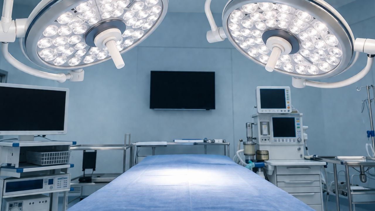
Surgeons need a bright light to perform a surgery. This is why theater rooms require modern LED surgery lights. The days of using yellowish halogen bulbs are over. Today, LED systems allow crisper, more natural color that resembles daylight. They prevent eyestrain in long surgeries, and their bright beam focuses on the area being operated on. We’ll list some of the best surgical lights for an operating room.
These lighting solutions are an integrated part of the surgical team. They give the surgeon a depth of field that keeps his or her focus sharp, whether he or she is performing a procedure on the surface of the body or a procedure deep within the body.
Touchless controls make it easy for the team to adjust the intensity and position the lighting without interrupting their sterile field.
What are Surgical Lights?
Surgical lights, also known as operating lights or surgical lightheads, are medical devices used during surgeries to illuminate a surgical site. These lights may look like expensive lamps, but they can perform specific tasks not possible with normal room lighting.
The role of the spotlight is to provide a high-intensity light stream that won’t create shadows or produce excessive heat. Here’s how they work and why they matter in an operating room.
Shadow Reduction
Surgical lights use several bulbs or clusters of LEDs, angled and spaced apart so that the beams overlap each other. If one hand blocks the path of light, the other fills it in. This wraparound method ensures the surgeon sees the tissue they are working on.
True Color Accuracy
Ordinary bulbs have a yellow or blue tint that can make organs look discolored. Surgical lights appear like natural daylight to help the team see the body’s real colors.
Heat Management
If an operating light reaches a certain temperature, it may dry out the tissues or cause discomfort for the surgical team during long procedures. Modern surgical lights use “cool light” produced by LEDs. LED lights beam so bright light without raising the temperature of the surgical area.
Light Intensity and Focus
Almost all of the lights allow the surgeon or nurse to adjust the brightness levels (in lux) based on the extent of the job at hand.
Reliable
Surgeons can snap on a cover of a sterilizer over these handles and easily change the location of the light without coming into contact with their gloves. The arms are balanced against the body of the light, so it remains exactly where it is supposed to be without moving.
Surgical Lights Configurations
- Ceiling-Mounted: Usually used in permanent operating rooms. They save floor space and allow the maximum range of motion.
- Mobile Lights: These lights are on wheels and plug into the wall. They are used for minor procedures or as backup light in a rescue situation.
- Headlamps: Some surgeons wear a light on their forehead so they can shine a beam exactly where they want, especially during heart or brain surgery.
Best Surgical Lights for Operating Rooms
Here are five of the best LED surgical lights on the market, reviewed by what they do and why they matter.
Steris Harmony iLED
A special feature about the Steris Harmony iLED is its optical precision. It uses a series of high-quality lenses that overlap to form a single solid column of light. This design ensures the consistency of light on the surgical site, even if the surgeon’s head or shoulder gets in the way.
Features: One of the features of the Harmony iLED is the individual control of the LEDs. Instead of a single large bulb, it uses dozens of individual LEDs with custom lenses. It also has “Spot Adjustable” technology, which allows the surgeon to adjust the size of the beam without moving the light head. It has a high Color Rendering Index (CRI), so tissues look exactly as they should under natural light.
Advantages: The light head has no moving parts. Many lights have mechanical focusing parts that can break down after a while. This is done electronically by the iLED. It also has a “Suspension Advantage” where the arms move smoothly and stay right where you pull them. It does not drift, which is a common problem with cheaper models.
Benefits: It decreases eye fatigue. The light is constant and similar to natural daylight, so the surgeon’s eyes do not need to adjust to bright or dim spots. In addition, LEDs produce virtually no infrared heat, so the surgical site stays cool. This prevents the tissue of the patient from drying out and also provides comfort to the surgical team during long, multi-hour procedures.
Dräger Polaris 600 Surgical lights
The Dräger Polaris 600 is built for the modern operating room. It’s a pared-down system that removes the clutter associated with older lighting rigs.
Features: This light has a sterile touch control handle. That means the surgeon can adjust the light intensity and the color temperature right from the sterile field without reaching for a wall panel or asking a circulating nurse. It also includes a “Mobile App Control” option that allows staff to pre-select lighting configurations for certain types of surgery.
Advantages: The Polaris 600 is quite light. For hospitals using laminar flow ventilation systems, the head’s thinness and aerodynamics are vital. The air that maintains the room’s sterility is not blocked. The ability to change the color temperature is an additional benefit.
Warm white and cold blue-white lights are switchable. When you need to differentiate between different types of tissue, such as blood vessels and nerves, this is useful.
Benefits: The enhanced workflow allows surgeons to adjust the light with the sterile handle. Also, it improves safety; the light is equipped with an HD camera system capable of recording or streaming surgeries for educational use without increasing overhead clutter. This feature is suitable for teaching hospitals.
Maquet Volista (Getinge)
Under the Getinge brand, Maquet is a popular name in operating room equipment. The Volista series is their leading lighting solution, specifically to address the issue of shadows in the surgical environment.
Features: The Volista has a “Shadow Management” mechanism that is patented. The light head’s many LEDs are configured to offer continuous lighting from various perspectives. Moreover, it has a “Boost Mode,” which is useful for deep-cavity surgeries where visibility is poor. It has a dedicated green ambient light mode for minimally invasive (laparoscopic) procedures.
Advantages: The Volista’s best feature is its ability to regulate shadows. The light “wraps” around obstructions to maintain illumination of the wound site even when many persons are bent over the patient. Another momentous benefit is the construction quality; the light head is sealed, which makes it easy to clean.
Benefits: Shadow management takes control once the light is positioned, removes the need for frequent human adjustments. For laparoscopic procedures, the green ambient light is a huge benefit because it doesn’t reflect on the video monitors. During keyhole procedures, it reduces errors.
Stryker Visum LED II
The Visum LED II is designed for deep-tissue visibility and dependability, and Stryker is a house-hold name in the surgical industry. It is a workhorse lamp made to withstand the demands of orthopedic suites and high-volume trauma hospitals.
Features: The brightness of 160,000 lux offered by the Visum LED II is at the upper end of what is safe and effective for human vision. Its large-diameter light heads generate a broad field of lighting. It has an exclusive heat-dissipation mechanism that draws heat out of the back of the light head and away from the LEDs.
Advantages: Some lights appear excellent at first glance, but as you go deeper into an incision, they become faint. Even at a distance, Stryker’s optics maintain a bright beam.
This implies that when the table is slanted or the surgeon switches from superficial to deep tissue surgery, the light doesn’t need to be changed as much.
Benefits: The surgical team doesn’t have to constantly divert their focus to reposition the light because of the depth of field. It offers a stable, daylight-like atmosphere that facilitates the identification of small structures, which is essential for complex orthopedic or vascular repairs.
Mindray HyLED 9 Series
The Mindray HyLED 9 Series is a high-performance light with an emphasis on “smart” technology and ergonomics. It is made to be among the thinnest light heads available, which sets it apart from the heavy lights of the past in both appearance and feel.
Features: The HyLED 9 has a special “cross-shaped” design. In addition to being aesthetically pleasing, this improves the flow of air around the light head. “Automatic Illumination Control” is another feature. This sensor-based technology automatically increases the power of the remaining LEDs to make up for the lost light when it detects that an object is obscuring an LED.
Advantages: It’s better for smaller hospitals. Surgeons can select a warmer light for the skin and a cooler light for bone and internal organs. There is no flickering and a seamless transition between these settings.
Advantages: The design’s aerodynamic form keeps the sterile air from being disturbed. It reduces the chances of surgical site infections. For the surgeon, it removes the “dimming” effect that occurs when they lean in for a closer examination.
How to Choose the Best Surgical Lighting for Your Operating Room
Test the Drift
A light is only beneficial if it remains where you place it. Poorly balanced suspension arms may cause the light to drift from its target, and that will require constant adjustment.
- Ease of movement: Can one nurse or technician move the light?
- Stability: Does it remain locked in position once you set it?
- Range of motion: Can the arm cover the full table, whether vertical or horizontal?
Integration
Several surgical lights have options to integrate HD or 4K cameras directly into the light head. Even if a camera isn’t necessary now, selecting a “camera-ready” model allows you to add video capabilities later for education, documentation, or remote consultations without replacing the whole system.
Easy to Sterilize
Choose light heads with smooth, seamless surfaces.
Avoid exposed screws or deep grooves, as these can harbor dust and bacteria.
Sterilizable handles: Make sure the center handle can be removed easily.
IP Ratings: Buy lights with a high Ingress Protection (IP) rating.
Best surgical lights Checklists
LED Technology: Offers longer lifespan and reduced heat compared to halogen.
Illumination Intensity (Lux): Typically ranges from 100,000 to 160,000 Lux.
Light Field Diameter: Should cover the surgical site adequately but be adjustable for smaller incisions.
Battery Backup: Confirm the system connects to the hospital’s emergency power or has an internal backup.
Read also: Top Endoscopy Systems for 2026 (High-definition Imaging Compared)
Final Thoughts on the Best Surgical Lights
Professional surgical lights should have clear, shadow-free vision without excessive heat production. High-quality LEDs are ideal because they maintain a cool environment, still deliver natural and precise color.
So if you’re looking for the best surgical lights, choose lighting systems with flexible, balanced arms. Reliability is essential; consistent and flicker-free performance reduces eye strain and enhances focus during long operations.
Hospitals are meant to be neat. If not well sterilized, people can contract infections from there. That’s where the best UV sterilization equipment’s for hospitals comes in. They have a special kind of light that kills germs without using chemicals. If you own a medical facility or are in the medical field, it’s vital that you know how these UV systems work and the list of equipment every hospital should have.
One problem is that it is hard to kill some germs. They can live on door handles, bedding, and medical instruments. Staff must always wash their hands and keep everything clean. Sometimes they get busy and skip a step.
In addition, visitors bring germs to the hospital from outside. Hospital staff face a daily challenge in infection control.
What is UV Sterilization?
UV is short for ultraviolet light. It’s a kind of light you don’t see with your naked eye. You hear it from sunscreen bottles, and you see it when it says you could be burning. The thing is, UV light also kills bacteria and viruses.
When UV light strikes the microbes, it damages their DNA or RNA. It’s like breaking the instructions that tell the germ how to act. If the “instructions” are broken, the germ can’t reproduce or do anything harmful.
How Does UV Sterilization Equipment’s Work?
It’s very simple. UV sterilizers contain a special bulb that produces UV-C light. That light is between 200 and 280 nanometers. When you turn it on, the light hits whatever is in it or walking by, and the germs are exposed. Depending on power, they are destroyed in seconds or minutes.
Some of these light-up devices use a closed box to illuminate items for a specific period. Others are mounted on walls or ceilings to project light over an entire area, targeting air or surfaces. The closed box version works more quickly because nothing obstructs the light. However, the room-mounted systems take longer but can cover a larger space.
UV Sterilization Equipment’s for Hospitals
Modern UV disinfection devices help healthcare facilities keep environments clean. The best UV sterilization equipment’s for hospitals today include mobile towers, storage cabinets, and air purification systems.
Mobile UVC Towers for Room and Surface Disinfection
These movable units remove the need for chemical cleaners. Hospitals can transport these towers to any location. Some of the best are:
STERO-360 (ALFAA UV)
This tower beams strong UV-C light throughout entire rooms that traditional cleaning can miss. Hospital cleaning teams can guide the unit into patient rooms, operating theaters, or waiting areas and start the disinfection cycle.
- Features: The STERO-360 uses powerful UV-C bulbs and smart sensors to detect if a room is occupied. It has a sturdy wheeled base that glides easily over hospital floors. A built-in timer lets staff adjust exposure times based on room size and infection guidelines. The unit also has protective shields to prevent accidental UV exposure during use.
- Uses: Hospitals use the STERO-360 in patient rooms after cleaning. Surgical teams use it to enhance final cleaning in operating theaters. Emergency departments use the unit for fast room changes during busy times. The tower is effective in isolation rooms where there is a higher risk of pathogens spreading.
- Advantages: The STERO-360 disinfects rooms in minutes instead of hours. This automatic system reduces the need for chemical disinfectants. Staff find it easy to use. The unit can reach under furniture, behind equipment, and into corners that manual cleaning can miss.
UV Sterilization Equipment’s for Hospitals: Ultroz UV-C Tower Plus
The Ultroz UV-C Tower Plus is among the best sterilized cleaning tools. It uses strong UV light to kill up to 99.9% of germs on surfaces.
- Features: The Ultroz Tower Plus has several high-powered UV-C lamps arranged to cover large areas effectively. It is mounted on a secure base with wheels that lock in place for safe operation. The tower comes with settings that allow users adjust cleaning cycles based on room size. Safety features like overload protection and automatic shutoff make it safe to use.
- Uses: Big patient rooms, ICUs, and procedure rooms benefit from the Tower Plus’s coverage. Hospital pharmacies use it to keep medication storage areas clean. Labs use it to clean workspaces between experiments. It is also useful in rehab centers and physical therapy areas to disinfect surfaces that are frequently touched.
- Advantages: Its powerful output requires fewer cleaning cycles for complete disinfection. The user-friendly controls help reduce mistakes and ensure consistent results. Built to last, it can handle the everyday demands of hospital environments. Its mobility allows it to be used in multiple areas within a hospital.
High-Power Mobile Trolley Sterilizer
These trolleys are popular due to their simple design and reliable performance, which is why health facilities go for it.
- Features: These mobile sterilizers have strong UV-C lights attached to height-adjustable carts. The wheels lock to keep the unit steady during use. The easy-to-use control panels enable quick start cycles without complicated programming. Many models have remote control features.
- Uses: Outpatient clinics use them between patient visits to keep exam rooms clean. Diagnostic imaging departments use these carts to disinfect surface areas before scanning.
- Advantages: It’s easy to maintain. Because they use a familiar trolley format, no special installation or changes to hospital infrastructure are needed. Staff can disinfect areas as required, instead of waiting for scheduled cleaning times.
UV Sterilization Cabinets for Small Equipment Disinfection
UV sterilization cabinets have safe enclosed spaces where hospitals can disinfect heat-sensitive instruments and small medical devices. Examples include:
Biobase UV Sterilization Equipment’s for Hospitals
Biobase have various sizes, like the popular 300L and 100L models, to meet different facility needs. It combines UV sterilization with temperature regulation, these units are versatile for sensitive uses.
- Features: Biobase cabinets have interiors designed to reflect UV light for full coverage. Shelves can be adjusted to customize the layout based on what’s being sterilized. Digital temperature controls help keep specific thermal conditions when needed. Sealed doors stop UV leaks when using it.
- Uses: Hospital labs use these cabinets to keep consumables and culture media sterile. Surgical departments store small tools in Biobase cabinets between uses. Pharmacy departments clean medication prep tools and measuring devices. Blood banks use the cabinets to clean equipment before important procedures.
- Advantages: The cabinets combine sterilization and temperature control to meet different hospital needs. Bigger models, like the 300L, can handle large amounts at once. The closed design keeps items clean until they’re used. They’re energy-efficient, so they help hospitals save on costs.
Medical UV Sterilizer Cabinets
Medical UV sterilizer cabinets are designed for hospitals to handle non-critical, heat-sensitive tools. These cabinets help healthcare facilities prepare items for patient use.
- Features: The polished aluminum interior has UV reflection for surface cleaning. The multiple UV bulbs ensure there is thorough coverage with no missed spots. Automatic timers make the operation easy, and interlock systems stop the operation if the cabinet is open.
- Uses: Outpatient clinics use these cabinets for stethoscopes, blood pressure cuffs, and other reusable exam tools. Dialysis centers clean the tubing and connections in them. Endoscopy units prepare scopes and accessories that can handle UV light but not high heat. Dermatology offices clean small instruments and treatment supplies.
- Advantages: The gentle UV method makes tools last longer than stronger chemical or heat methods. Fast cycles allow quick processing during busy clinic times. They are compact, fit easily in exam rooms and nursing stations. Simple to use, and they require minimal staff training.
Air Sterilizers for Airborne Pathogen Control
Air sterilizers tackle the unseen threat of airborne germs in hospital air systems and public spaces. These machines cut down the spread of respiratory infections. Modern UV air sterilizers kill microorganisms without making harmful byproducts or ozone.
Ultroz UV-C Air Sterilizer
The Ultroz UV-C Air Sterilizer is designed to tackle airborne germs in busy hospital hallways and patient rooms. This air purification device gets rid of mold spores, bacteria, and viruses as they pass through its sterilization chamber. Hospitals place these units where continuous air treatment is mostly beneficial.
- Features: The Ultroz system comes with strong UV-C lights set in the air path to ensure maximum germ exposure. Efficient fans move large amounts of air through the chamber. Filters catch particles before they reach the UV lights.
- Uses: Hospital corridors use ongoing air treatment between patient rooms. In ER waiting areas, these purifiers help protect vulnerable patients. Intensive care units use the devices to lower infection risks for seriously ill patients. Operating room prep areas and recovery rooms need them for quality air.
- Advantages: Non-stop operation means constant protection instead of occasional treatment. The systems cut down on airborne germs without using chemicals or noise. Setup is easy and doesn’t require significant changes to hospital infrastructure. Low running costs make them suitable for long-term use.
SOTO UV360 Portable Purifier
The SOTO UV360 Portable Purifier are in a portable form. Medical facilities appreciate the ability to move the purifier wherever air quality is a priority.
- Features: The UV360 has a 360-degree air intake that draws contaminated air from every angle into its sterilization chamber. Several UV-C bulbs ensure germs are destroyed for consistent results. Wheels make it easy to move as needed. Smart sensors keep track of air quality and adjust performance automatically.
- Uses: The UV360 is used in administrative areas to keep the air healthier. Mobile health units also take advantage of their portable sterilizing features. In hospitals, isolation rooms include these purifiers to enhance air treatment. Dental offices and outpatient surgical centers rely on them to improve air quality in treatment rooms.
- Advantages: Because it is portable, a single unit can be moved to different areas instead of installing one in each place. Built to handle tough hospital conditions, its commercial-grade design is durable. It runs automatically, so staff don’t need to spend time checking on it. Its small size makes it suitable for areas where larger systems wouldn’t fit.
UV Sterilization Equipment’s for Hospitals Checklists
Wavelength Effectiveness
UV sterilization works because certain wavelengths of ultraviolet light break apart the DNA and RNA of germs, bacteria, and viruses, stopping them from multiplying and causing infections. The ideal wavelength you need is 254 nanometers, which is part of the UV-C category and is most effective at killing germs.
When checking out equipment, look at the UV-C output, which is usually mentioned in nanometers by manufacturers. Make sure that the lamp provides a strong, consistent output at the 254nm wavelength. Ask the manufacturer if they offer guarantees on wavelength stability.
Also, find out if the system uses mercury-based lamps or newer UV-LED technology. Both can operate at the right wavelength, but they differ in performance. Mercury lamps are cheaper but need frequent replacements, while UV-LED systems cost more but last longer and reach full power instantly without warming up.
UV Intensity
The success of UV sterilization depends not only on the presence of UV light but also on the amount that reaches the surfaces. This is measured by the dose, expressed in millijoules per square centimeter (mJ/cm²). Intensity indicates the strength of the light at its source, while the dose considers both intensity and exposure time to determine if sufficient germ-killing power reaches the target areas.
Different germs need specific doses to be completely deactivated. In healthcare settings, most bacteria and viruses require a dose between 20 and 100 mJ/cm² to achieve 99.9% elimination. Ensure your equipment can deliver the necessary dose for the pathogens present in your environment.
Room Coverage
A UV sterilization unit may cover 1,000 square feet, but you need to know how light spreads in a room. UV light moves in straight lines and doesn’t curve around corners. Placing a unit in the middle of a room ensures good coverage directly in front and above it, but areas behind furniture, tables, and corners might get little to no UV exposure.
The way UV light covers a room is important for effective disinfection. Some units emit light in a 360-degree pattern from a central location. Others might project light in shapes like cones or directional beams or use multiple bulbs aimed at various spots.
Power Requirement
Most commercial UV sterilizers work with standard 120V or 240V electrical systems, depending on size and power. Make sure your facility’s electrical system can handle the units, especially if they require special circuits due to high output.
Besides basic power compatibility, consider energy efficiency ratings. Efficient units provide the same disinfecting power while using less electricity. As these machines may operate multiple times daily, every saved watt results in significant long-term savings.
Some systems have power-saving modes or smart operations that adjust intensity based on room size or occupancy detection.
Read also: Wireless Patient Monitoring Systems for Operating Rooms
Final Thoughts: UV Sterilization Equipment’s for Hospitals
UV sterilization equipment’s for hospitals kill DNA and RNA of bacteria through its targeted light. You can you use for water treatment, air purification, and surfaces we touch daily.
As more people learn about these tools and technologies, they will be found in more homes and public places. We can stay healthy in clean spaces, and UV sterilization provides an easy way to protect ourselves in and outside the hospital.
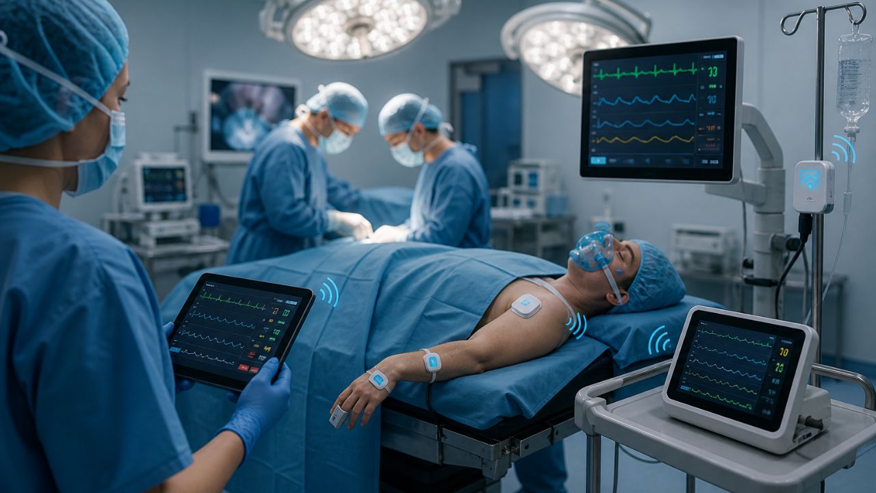
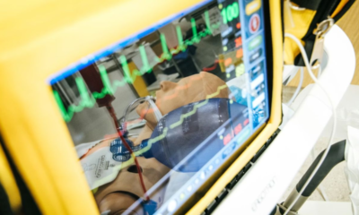
A cable from traditional patient monitors causes tripping hazards and sometimes makes it hard for the surgical team to access vital signs. What if we remove this clutter and create a cleaner, safer surgical space? That’s where wireless patient monitoring systems step in. These devices eliminate clutter and enable surgeons and nurses to focus fully on the patient. Wireless monitors track vital signs and send alerts without restricting movement, making procedures easier.
The reason for adopting this technology is safety. Tripping over cords in a busy operating room is not just irritating; it can be dangerous. A cord might dislodge an IV or disconnect a central monitor. However, a wireless system removes this danger.
Also, these devices use encryption to safeguard patient information, ensuring that signals are only received between the patient and their doctor.
What are Wireless Patient Monitoring Systems?
Wireless patient monitoring systems remove the need for physical cables. Their main function is to continuously track a patient’s vital signs during surgery, achieving this without requiring direct cable connections.
How do these wireless systems work?
Small sensors, which are easy to forget about, stick onto the patient’s skin, similar to what you’ve seen in hospitals. These sensors don’t have wires coming out of them; instead, they contain small built-in transmitters. These transmitters send important data wirelessly over a secure medical network to a central station.
This station then displays all the vital signs clearly on a screen in the operating room, and sometimes even in another control room.
Difficulties with Traditional Wired Monitoring
If you’ve ever visited a cardiac or intensive care unit, you might have noticed the tangled web of wires connecting patients to their beds. Although these wired systems have been lifesavers for years, they come with downsides that can make things tough for both patients and nurses.
Limited Mobility
Patients are encouraged to sit up, walk to the bathroom, or stroll down the hallway. However, wired monitoring can feel restrictive. When connected to a wall-based monitor, even going to the bathroom can be difficult.
Nurses might need to come in, disconnect the wires, and attach them to a portable unit. This process takes time, and many patients prefer staying in bed rather than dealing with the hassle of wires.
Safety Concerns
Wires can twist under the bed rails. This can cause extra strain on the mattress frame, tripping hazards, or false alarms. Any of these saety concerns can affect either the patient or the caregivers.
Challenges with Hygiene
Thick, ribbed plastic cables can collect germs easily. Tiny viruses or bacteria can sneak into the crevices of these cables, unlike on a smooth, wireless sensor.
Discomfort Concerns
Hard plastic connectors dig into the back or arms, causing skin irritation. For patients who need rest to recover, it’s tough to get quality sleep with wired monitoring.
Cost of Maintenance
Cables frequently break, and replacing them is not cheap. Hospitals spend thousands of dollars each year dealing with worn-out leads and damaged connectors.
Wireless Patient Monitoring Systems Benefits
Wireless patient monitoring systems are revolutionizing hospitals. Here’s why it matters:
Better Flexibility
With wireless systems, surgical teams get more flexibility. With less bulky equipment, it improves comfort and efficiency for everyone involved.
Enhanced Safety
Infection control is vital in healthcare. Traditional monitors with several wires and connectors are difficult to clean. Wireless systems solve these issues by using flat, simple, patch-like sensors placed on patients’ skin. These smooth surfaces don’t have places where bacteria can hide.
Keeps Patient Care Consistent
When patients transition from before surgery to the operating room and then to recovery, they are often moved between different beds and rooms. In the past, this meant nurses had to manually unplug and reattach all monitoring equipment, which could lead to losing important data.
A small sensor stays with the patient and sends data to the monitors without interruption. This guarantees that the medical team has a complete and accurate record of the patient’s condition, capturing all crucial information during these moves.
Advances Driving Change
The process isn’t just about getting rid of wires. It involves a complete transformation powered by new technology, such as:
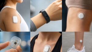
Skin-Based Technology: Wearable Biosensors
These small, sticky patches go on a patient’s chest and contain various tiny sensors. Whether the patient is moving, resting, or walking, these gadgets stay active. They constantly check vital signs like:
- ECG: Monitors heart rhythm for any irregularities.
- SpO2: Measures the oxygen level in the blood.
- Respiration: Counts the number of breaths per minute.
- Temperature: Checks for possible fevers.
Being both wireless and comfortable, these patches allow patients to move around freely, enable doctors to monitor the physiological data of their patients.
Reliable Data Transmission
The data from the biosensors needs to efficiently reach the nurses’ station, which is where connectivity is vital. It’s the unseen route that carries the information. The system uses smart, energy-saving options to maintain a constant and reliable signal:
- Bluetooth Low Energy (BLE): This short-distance method lets the patch talk to a small hub in the patient’s room, using very little power.
- Wi-Fi: The room hub then uses the hospital’s secure Wi-Fi network to send the data to the building and into the cloud.
- Medical Body Area Networks (MBAN): A secure path set aside for medical devices, avoiding interference to ensure vital data is always sent.
EMR Integration
Data doesn’t do much good if it’s kept separate. Its true value comes when this wireless information directly links with the patient’s electronic medical record (EMR or EHR). This removes the need for manual data entry and waiting for a nurse to update readings.
The system continually refreshes the patient’s digital record, giving the medical team a real-time view of the patient’s condition. This helps them spot trends and react quickly to changes, turning raw data into useful information.
Cybersecurity
With sensitive health data being sent around, is it secure? Absolutely. In a wireless hospital setting, keeping patient privacy intact is vital. Systems use strong encryption like a secret code, so even if the data is intercepted, it can’t be read.
They also use secure networks, enforce strict access controls (making sure only authorized individuals can access the data), and keep a watchful eye to detect any potential threats. It’s all about protecting health information digitally.
The combination of wearables, connectivity, intelligent software, and strong security is changing patient care from being static to a continuous, dynamic process.
Technologies & Systems
This setup gives medical teams an ongoing, real-time view of a patient’s health. Let’s look at two key technologies that makes this progress possible.
Wearable Sensors & Patches
They are small, lightweight sensors that look like an adhesive patch placed on the chest. It performs tasks previously handled by multiple traditional machines. Products like the Sensium Vitals Patch and the VitalPatch act as efficient and compact health monitors.
Once attached to the skin, they quietly measure various vital signs, such as heart rate, breathing rate, oxygen levels, and body temperature. In addition, the Isansys Lifecare Patient Status Engine gathers this data and securely transmits it to a dashboard to provide continuous health updates rather than periodic ones.
Cable-Lite Systems
Sometimes, a bedside monitor is still necessary, but however, Cable-Lite” systems solve this problem. Advances like the Mindray BeneVision V Series cut down on cable use. It improves both safety and comfort in healthcare settings.
This makes it easier and quicker to move patients between departments and reduces the chance of accidentally unplugging critical equipment. These systems offer the strong monitoring features of traditional devices with the sleek, patient-friendly design of modern technology.
Common Wireless Components
ECG & Heart Rate
Patients can wear a small, nearly invisible patch on their chest. This patch keeps a constant watch on the heart’s electrical activity, detects any irregular patterns like skipped beats or rapid rhythms as they happen. The essential data is sent wirelessly to the healthcare team, without confining the patient to bed.
Pulse Oximetry (SpO₂)
The tiny clip placed on a patient’s finger is now available in a wireless model. It measures blood oxygen levels and sends the data directly to the central monitoring system, eliminating the need for nurses to check manually. It ensures that the body receives sufficient oxygen, needed for patients with respiratory issues.
Blood Pressure
Wireless blood pressure cuffs have improved on their earlier versions. They can be programmed to take measurements regularly throughout the day and night. Each reading is sent immediately to the patient’s electronic record, creating a comprehensive chart of blood pressure fluctuations.
Respiration Rate
It’s important to track how many breaths a patient takes each minute, but manually counting them is not possible. Wireless systems have tackled this issue with smart sensors embedded in a comfortable vest or a soft chest strap. These sensors detect the small movements of the chest with each breath. If the breathing rate becomes too slow, too fast, or stops, the system alerts the medical staff.
Temperature
Instead of using a thermometer all the time, a small wearable patch can be attached to the skin to continuously track body temperature. It can monitor even the smallest changes in fever or temperature drops.
Read also: Robotic Knee Replacement Surgery: The Future of Joint Relief
Final Thoughts
Introducing wireless monitoring into operating rooms is a major upgrade. No more tangled cables, just clear, real-time information that makes patient care safer and easier.
This allows the surgical team to focus on the patient without interruptions. Basically, it’s not just about having cool gadgets; it’s about genuinely improving people’s lives and guaranteeing a safe surgical experience.
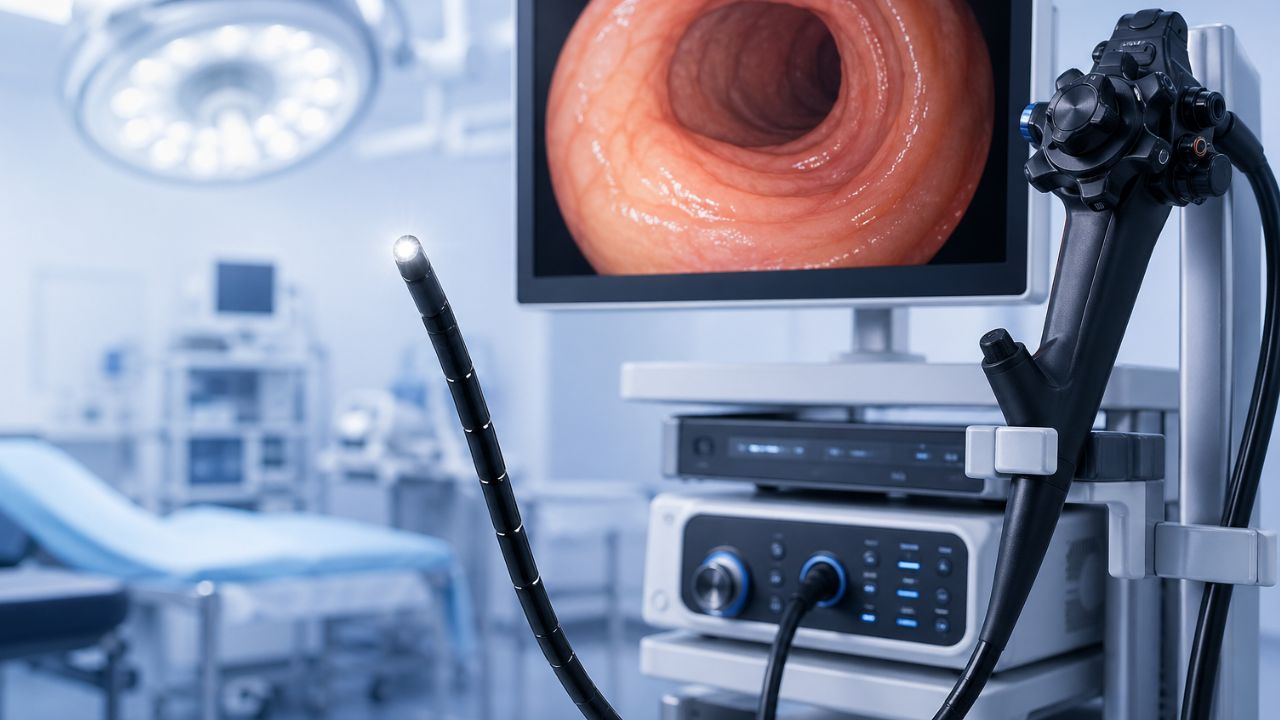
Doctors can look inside your body, take pictures or videos of your organs and tissues, and then use those images to detect, and treat health problems without having to do surgery. This is what endoscopy is like today. We’ll discuss the top endoscopy systems on the market in 2026.
Olympus and Fujifilm are among the best companies. Their equipment turns small textures into clear images. Olympus is better because it uses special light filters that make small blood vessels stand out in green or red. This makes it easier to find flat growths that are usually hard to see.
Fujifilm works differently. They use four LED bulbs to mix light in a way that shows the real color of internal organs without any delay. At the same time, the current Pentax models focus on digital sharpening, which keeps videos clear even when the camera moves quickly.
These systems improve standard video as they add smart overlays that show areas of concern in real time. The upgrade from rough video to 4K clarity allows doctors to detect any abnormalities accurately.
What is an Endoscopy System?
An endoscope is a long, thin, flexible tube with a small camera and a bright light at the end. The doctor starts the process by putting the tube through a natural opening, like the mouth or nose, or through a small cut in the skin. The camera records high-definition video of the inside of the body as the tube moves through it.
This video goes back through the tube and appears on a big screen in the exam room. The doctor watches this live feed to look for signs of swelling, redness, blockages, or abnormal growths.
Modern systems provide more than just a view. They have extra channels, small holes in the tube that allow doctors to slide tiny tools down to the tip of the tube. With these tools, a doctor can cut off a small piece of tissue to run a test, stop bleeding, or even remove a growth on the spot.
Interestingly, these systems use natural openings or small cuts, which means they are less painful and require less time to recover than traditional procedures.
What are the three types of Endoscopy?
Doctors put endoscopies into groups based on what part of the body they look at. Each uses a thin, flexible tube with a light and a camera to view inside the human organs. The types are:
Upper Endoscopy
This test looks at your digestive tract from the throat to the top of the small intestine. A doctor uses a scope to check for ulcers, inflammation, or blockages in your esophagus, stomach, and the first part of your duodenum. If you have heartburn, stomach pain, or trouble swallowing, they may perform this test.
Colonoscopy
The doctor passes the scope through the rectum and looks for polyps, tumors, or areas of bleeding. This test is used for colorectal cancer screening.
Bronchoscopy
The doctor places the scope through your nose or mouth and into your lungs. The scope examines your windpipe and bronchial tubes to know exactly the cause of chronic cough, identify a blockage, or obtain a tissue sample if there is symptoms of lung infection or mass.
What Diseases can be Detected by an Endoscopy Systems?

When asked, “What diseases can be detected by endoscopy?” the answer includes several conditions that affect your digestive system.
Examination by an experienced, qualified healthcare professional allows the doctor to examine the stomach to find the cause of persistent stomach pain, bleeding, or difficulty swallowing. During this examination, the doctor can look for:
Ulcers: The camera reveals open sores on the lining of the stomach or the start of the small intestine. These cause burning pain and can lead to internal bleeding if the fails to treat it.
Inflammation: If your stomach or esophagus appears red, swollen, or irritated, the doctor notes conditions like gastritis or esophagitis. This often explains why you feel constant heartburn or nausea.
Gastroesophageal Reflux Disease (GERD): In this case, the scope will check if there is damage to the esophagus from stomach acid moving in the wrong direction.
Celiac Disease: Doctors may remove a small piece of tissue to see if your immune system is causing damage to your small intestine after you eat gluten.
Growths or Polyps: The camera identifies abnormal cells and can be used to take them out before they have a chance to develop into something more serious.
Cancer: If an area of concern appears abnormal, the doctor performs a small biopsy. Lab analyses of this tissue will confirm whether or not you have cancer of the esophagus, stomach, or small bowel.
Narrowing or Blockages: At times, scarring or tumors can cause parts of the digestive tract to become too narrow, making it difficult for food to pass. Endoscopy helps locate exactly where these blockages are.
Endoscopy Systems for Modern Hospitals: The Best Equipment’s
The equipment you choose can affect the accuracy of diagnostics. Below are the top five endoscopy systems on the market.
Olympus EVIS X1
The Olympus EVIS X1 is a top pick for GI departments that want the latest diagnostic technology. This system focuses on early detection and is user-friendly.
- Feature: It has Extended Depth of Field (EDOF) and Red Dichromatic Imaging (RDI) technologies.
- Advantage: EDOF ensures that both close and distant tissues are in focus without needing adjustments, while RDI helps visualize deep blood vessels more clearly.
- Benefit: Doctors can perform procedures faster and spot hidden lesions or bleeding more accurately.
Fujifilm Eluxeo 7000 Series
The Fujifilm Eluxeo system is known for its exceptional lighting capabilities. It’s a versatile tool suitable for both regular screenings and complicated treatments.
- Feature: It uses Multi-Light Technology with 4-LED illumination, plus modes like Blue Light Imaging (BLI) and Linked Color Imaging (LCI).
- Advantage: These modes make the contrast between healthy and abnormal tissues clearer, showcasing mucosal patterns distinctly.
- Benefit: This clarity helps doctors identify polyps and tumors immediately. So it reduces unnecessary biopsies and improves patient care.
Pentax Medical OPTIVISTA EPK-i7010
The Pentax OPTIVISTA combines high-definition imaging with digital enhancements. It’s praised for its easy-to-use interface and fast image processing.
Feature: It includes i-SCAN technology and Twin Mode display.
Advantage: i-SCAN applies digital filters to enhance surface textures in real-time, while TwinMode lets doctors view both original and enhanced images side-by-side.
Benefit: Very useful for mapping and detecting lesion margins. It ensures complete treatment of the affected area.
Stryker 1688 AIM 4K Platform
While most systems target GI procedures, Stryker excels in surgical and multi-specialty endoscopy. It’s essential for laparoscopic and orthopedic surgeries.
- Feature: Delivers 4K quality along with SPY fluorescence imaging.
- Advantage: The 4K quality offers four times more detail than typical systems, and SPY mode enables surgeons to observe real-time blood flow in tissues.
- Benefit: This helps surgeons work with greater clarity and precision, reducing complications and speeding up patient recovery after surgery.
Ambu aScope (Single-Use Platform)
For hospitals focused on infection control and quick responses, Ambu leads in disposable endoscopes. It’s especially useful in ICUs and emergency settings.
- Feature: A fully sterile, single-use endoscope that connects to a portable high-def monitor.
- Advantage: It eliminates the need for complicated cleaning processes and equipment transport.
- Benefit: The risk of patient cross-contamination is removed, along with the costs for scope repairs and cleaning supplies.
How to Choose Endoscopy Systems for your Hospital
For patient your clinical workflows, consider these tips:
Superior Image Quality
The main job of an endoscopy system is to provide clear images. Surgeons and doctors depend on precise visuals to find issues or carry out delicate procedures. Focus on systems that provide:
- High-Definition (HD) Resolution: Sensors should deliver clear, noise-free images.
- Advanced Lighting: Get systems that use special light modes, like narrow-band imaging.
- Natural Colors: The endoscopy camera system should represent tissue colors to differentiate healthy and diseased areas.
Comfort and User-Friendliness
If the endoscope is hard to control, it can make long shifts challenging. Evaluate the equipment for:
- Easy-to-Use Handles: Controls should be comfortable and respond to small movements.
- Lightweight Design: Lighter scopes help reduce the risk of strain for medical staff.
- Simple Controls: An easy control panel helps staff adjust settings without going through complicated menus.
Durability
- Durability: Opt for scopes with strong parts and materials that can withstand wear.
- Reliable Reprocessing: Ensure the system can endure your hospital’s cleaning process without frequent issues.
- Repair Data: Investigate the “mean time between failures” (MTBF) for models.
Compatibility and Upgradability
Before you buy, confirm that the new system will fit with your existing setup.
- Monitor Matching: Will the new system work with your current surgical displays?
- Software Compatibility: Can the system save images and videos directly in your Electronic Health Records (EHR) or Picture Archiving System (PACS)?
- Room to Grow: Can you add new features or updates later, or will you need a new system when technology improves?
Vendor Assistance and Training
A sophisticated endoscopy system is effective only if your staff know how to use it. Good vendor support is as necessary as the equipment.
- Loaner Options: If a scope needs repair, does the vendor offer a temporary replacement to maintain your surgical schedule?
- Quick Support: Check how promptly the manufacturer’s service team addresses problems when a system fails.
Total Cost of Ownership (TCO)
Consider what it will cost to maintain the system over the next five to ten years. This should include:
- Energy Usage: Modern LED systems use less electricity than old halogen lights.
Top Endoscopy Systems (The Comparison)
| System Model | Primary Strength | Key Image Tech | Best For |
| Olympus EVIS X1 | High-contrast detail | TXI & RDI modes | Detecting bleeding & early lesions |
| Fujifilm ELUXEO 7000 | Artificial lighting | Multi-Light Technology | Identifying mucosal changes |
| Pentax OPTIVISTA EPK-i7010 | Digital zoom & clarity | i-scan OE | Narrow-band tissue analysis |
| Stryker 1688 AIM | Versatile integration | 4K fluorescence | Complex surgical procedures |
System Breakdowns:
Olympus EVIS X1
This system is great at detecting hidden problems. Its Texture and Color Enhancement Imaging (TXI) brings out slight changes in tissue texture. The Red Dichromatic Imaging (RDI) mode helps doctors identify bleeding sources.
Fujifilm ELUXEO 7000
Fujifilm stands out with its special four-LED light source that creates different viewing modes. It can quickly change light wavelengths, letting doctors switch from regular white light to enhanced vascular views just by pressing a button.
Pentax OPTIVISTA EPK-i7010
Pentax highlights the power of digital enhancement. Its i-scan Optical Enhancement (OE) technology uses light filters to make the edges of polyps and tumors clearer. This system provides sharp, high-definition images, aiding doctors in making quicker decisions during screenings.
Stryker 1688 AIM
Stryker designs this platform for today’s operating rooms. It offers 4K resolution and works well with other surgical tools. The AIM platform supports fluorescence imaging, giving surgeons instant visual feedback on blood flow and tissue health during surgeries.
Read also: Laparoscopy Equipment’s: The Top 5 to Buy for Your Hospital
Final Thoughts
The endoscopy systems set a new benchmark for clinical efficiency and patient safety. Its advanced imaging provides clear views, enabling doctors to identify issues and carry out smooth procedures faster. Hospitals investing in this technology enhance both diagnostic precision and workflow efficiency.
-
 Diagnostic & Hospital Equipments4 months ago
What Are Medical Devices? Types and Uses
-
 Uncategorized4 months ago
6 Best Heatmap Plugins – I Test, Review and Compare
-
 AI in Health care4 months ago
Remote Patient Monitoring in Modern Healthcare
-

 AI in Health care2 months ago
Digital X-ray vs. Film X-ray: Which is better?
-

 Review and guides4 months ago
CPAP Machines Buying Guide: The Best Models Doctors Don’t Recommend
-
 AI in Health care4 months ago
AI in Medical Imaging: What is Coming Next
-
 Diagnostic & Hospital Equipments2 months ago
DEXA Bone Density Scanning: What You Need to Know About Bone Health
-
 Diagnostic & Hospital Equipments1 month ago
PET Scan Machines: Understanding the Technology, and Usage