Diagnostic & Hospital Equipments
Best Surgical Tables in 2026 ( Uses, Features and Benefits
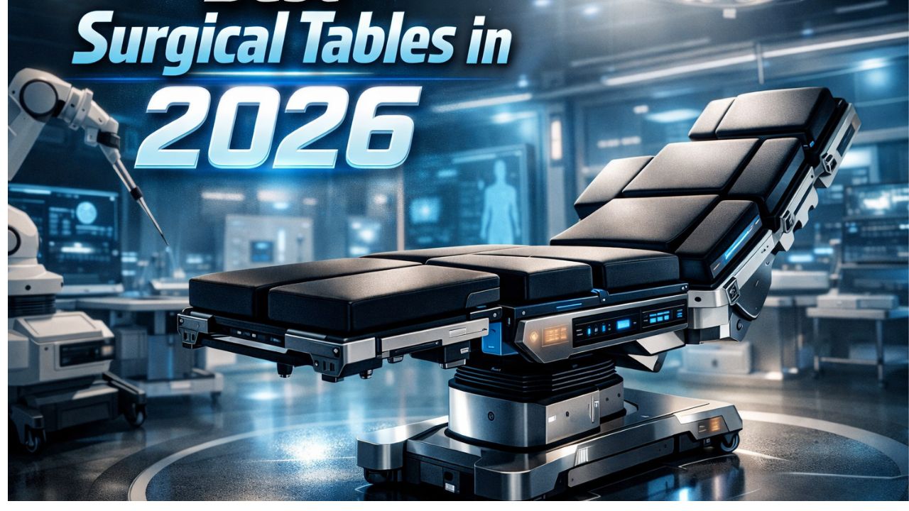
A surgical table is an advanced medical device that patients lie on during surgery. Robotic assistance, real-time imaging, and data-driven ergonomic design, however, has made it difficult to select the appropriate surgical tables.
Hospitals don’t buy medical equipment’s because of marketing claims from social media influencers; they go for the benefit it offers, capacity, and sometimes availability. As the need for surgical operations is increasing, the best healthcare facilities can do is to get devices that are clinically tested and patient-friendly.
What is a Surgical Table?
An operating table, also called a surgical table, is not a regular flat-surfaced table, it functions as an adaptable work platform, that requires stability when performing surgical operations.
It ensures proper patient positioning and allow surgeons to move freely while carrying out a surgery. These tables have modular designs and movable parts that enable precise adjustments. This includes changes in height, side-to-side tilt, and Trendelenburg or reverse Trendelenburg positions for different types of surgeries.
Modern tables are classified according to their clinical use, such as radiolucent tables made especially to work with X-rays and C-arm imaging, orthopedic, neurological, or general surgery. Pressure-relieving cushioning to stop skin deterioration and a variety of attachments, such as arm boards, stirrups, and headrests.
Many are driven by electro-hydraulic systems, which allow the surgical team to make seamless adjustments using a remote control.
What Defines a “Best-in-Class” Table?
We must know the three foundations of surgical technology before we discuss the best models. They are:
- AI-Driven: To reduce the danger of pressure ulcers and nerve damage during lengthy procedures, modern tables now use weight-distribution sensors to recommend the ideal Trendelenburg angle.
- Total Radiolucency: As a hybrid becomes more common, 360-degree carbon-fiber tops are used because they allow intraoperative fluoroscopy and CT without moving the patient.
- Robotic Syncing: To preserve the balance of the surgical field, some facilities integrate robotic platforms (such as Da Vinci or Hugo) into their operating tables.
Surgical Tables and their Uses
Maquet (Getinge)
Maquet, a Getinge Group brand, is always at the top of the list when it comes to the best surgical tables available on the market. With a history spanning more than 180 years, Maquet is now under the Getinge umbrella. From the mobile Maquet Meera to the modular Maquet Magnus, their surgical tables can handle both simple outpatient operations and multidisciplinary surgeries.
Uses:
General Surgery: Offers the steady support and comfort positioning required for procedures on the abdomen, gastrointestinal tract and other soft tissue areas.
Orthopedics and Traumatology: Maquet tables provide the precise traction and positioning for hip replacements, fracture fixations, and spinal alignments through their customized extensions.
Cardiovascular and Hybrid: These tables deliver minimally invasive heart and vascular procedures and can be integrated with advanced imaging systems (such as C-arms).
Neurosurgery: For delicate cranial procedures, it is compatible with skull clamps and specialty headrests.
Bariatric surgery: Helps to sustain high weight capacities.
Features of Maquet (Getinge) Tables
- Modular tabletop system
- Extreme Articulation and Positioning
- Superior Radiolucency
- Advanced Ergonomics and Intuitive Controls
- Auto-Drive)
Benefits:
Patient-friendly
The table’s ergonomic structure has specialized pressure-distributing pads (SFC padding) that reduce nerve injuries during extended procedures. Secure locking mechanisms maintain patient stability, even when the table is tilted to extreme positions.
Workflow Efficiency
Modular “transporter” components enable patients to be positioned outside the operating room before being moved directly onto the table base. This streamlines room transitions, cutting down turnover time and supporting a higher volume of surgeries each day.
Future-Proofing the OR
Maquet tables can integrate seamlessly with advanced 3D imaging technologies. Also, they can adapt as medical innovations progress.
Maquet Models to Consider
- Maquet Magnus
- Maquet Meera
- Maquet Alphamaquet
Skytron
Because of its user-friendly concept, Skytron is a favorite among surgeons and nurses. In terms of total-room maneuverability, the Skytron 3600 and 3500 Series are the top choices.
But this consistent reputation? Is it a strong brand presence, or is there a difference in how these tables function?
Unlike some manufacturers that concentrate on a single specialty, Skytron creates adaptable tables to serve multi-purpose solutions across hospital settings.
Uses for Skytron Surgical Tables
You’ll find them used across several medical specialties, such as:
- General Surgery: Handles routine appendectomies to complex abdominal procedures.
- Bariatric Surgery: Skytron has high weight-bearing capabilities.
- Orthopedics: Their tables provide the necessary traction and severe angles for knee and hip replacements.
- Cardiovascular & Neurology: They are ideal for operations that requires C-arm access because of their compatibility with medical imaging devices.
- Urology and Gynecology: Offer smooth, and accurate adjustments for tilting and lithotomy positions, enhancing procedural precision.
Features
- 270° to 360° Tabletop Rotation
- Industry-Leading Weight Capacity (Many models can support up to 1,000 lbs of lift and 800 lbs)
- Massive Top Slide
- Low-Profile Base
Benefits:
Patient Safety
Has an auto-beach chair with positioning and locking mechanisms that reduce the chance of the patient’s movement or slipping during procedures. These tables maintain steadiness, even when tilted too high.
Comfort
During lengthy microsurgeries when the table is lowered, or when working at elevated levels, it reduces back strain on tall surgeons.
Durable
Constructed from high-grade materials, these tables tolerate frequent exposure to chemicals and the constant use in high-volume trauma environments.
Efficient
Since a single Skytron table can replace three specialized units, hospitals benefit from reduced equipment needs, lower storage demands, and cheap costs to manage multiple devices.
Steris
While Skytron emphasizes mobility, Steris focuses on smart functionality. The Steris 5085 and 4085 models are the most dependable, and they can be integrated with hospital electronic medical record (EMR) systems.
These tables are efficient, whether in a small outpatient surgery center or a large, high-volume Level 1 trauma facility.
Uses for STERIS Surgical Tables:
General and Bariatric Surgery
Many models can carry patients weighing 1,000 lbs. These strengths are essential for bariatric surgeries, where safety and secure positioning are paramount.
Orthopedic and Spinal Surgeries
Precision in patient positioning is essential during orthopedic and spine operations. STERIS tables are compatible with a variety of specialized attachments that enable accurate limb alignment or facilitate prone positioning, such as tucking.
Their seamless integration with carbon fiber components also gives surgeons unobstructed access to the skeletal area, making these tables a preferred choice in complex bone and spine cases.
Advanced C-Arm Imaging
With the growing use of minimally invasive techniques, high-quality imaging has become important. STERIS tables have extensive radiolucent sections that allow clear X-ray imaging and efficient C-arm use. This design removes the need to reposition patients or deal with metal interference.
Features:
- Auto-Limit Sensors
- Exceptional Articulation.
- Intuitive Hand Controls
- Radiolucent Tabletops
- Self-Leveling Floor Locks
- Modular Design
The Benefits:
Lowers the Chance of Falls
Intraoperative falls and pressure injuries are less likely to occur when there is stability. High-quality pressure-reduction pads are used in conjunction with STERIS tables to distribute a patient’s weight and prevent bedsores, which can develop during extended, multi-hour marathons.
Enhanced Staff Ergonomics
The tallest surgeons and the lowest technicians may operate in a position that doesn’t damage their backs due to their height adjustments.
Berchtold Operon (Stryker)
The Berchtold Operon (D-Series), now part of the Stryker ecosystem, has established itself as one of the leading options for bariatric and orthopedic trauma procedures. German engineering and American innovation are combined in models such as the D850, D820, and D760. These are multipurpose surgical tables with sliding tops that may be used for nearly any operation.
Uses:
- General Operation: Equipped with specific accessories, it can be used for different types of surgeries.
- Orthopedics: Suitable for hip replacement.
- Cardiovascular & Urology: Its radiolucent design allows uninterrupted C-arm imaging, which is why it is ideal for image-guided interventions.
- Bariatric Surgery: With a high weight limit, it offers secure and stable support for patients needing additional capacity.
- Neurosurgery: During microsurgery, the Operon’s stability guarantees that delicate head-frame attachments stay stable.
Features:
- Massive Weight Capacity: The Operon D860 can support weights up to 1,250 lbs in a level position and 1,000 lbs through a full range of motion.
- Carbon Fiber Integration
- Sliding Top
- Ergonomic Hand Control
- Superior Articulation
The Benefits:
Sturdy
Even during high-torque orthopedic surgery, the table won’t move because of the floor-locking system. In addition, the pressure-relieving pads lessen the chance of “bedsores” during extended table time.
Dependable
This table offers the foundation required for heavy-duty surgical workflows for facilities that are seeing a rise in bariatric patients or those who have invested in the Stryker iSuite (the integrated digital operating room).
Mizuho OSI
When it comes to patient positioning, especially for orthopedic and spine surgeries, Mizuho OSI is still the undisputed choice. Their tables address the physical constraints that surgeons encounter, whether they are used for sophisticated imaging, orthopedic trauma, or spinal surgery. The ProAxis and Trios systems are top models.
Uses and Clinical Applications:
Spine Surgery: For spine surgeries, the Jackson Table is the industry standard. It enables advanced prone placement, lowers intra-abdominal pressure and enhances hemodynamics.
Orthopedic Trauma: Their platforms can handle the traction needed for tibia and femur fractures. It gives surgeons the stability they need to achieve ideal alignment.
Comprehensive Imaging: In settings where intraoperative C-arm imaging is required, these tables are essential due to their open frame design.
Features:
- Software-Controlled Positioning: The ProAxis table features motorized joints that resemble natural human movement.
- Radiolucent Dual-Column Structure: The table’s design creates unrestricted 360-degree C-arm rotation around the spine or limbs.
Benefits:
Improved Imaging Precision: Since the table does not interfere with X-rays, clearer images can often be obtained using lower radiation levels, minimizing exposure for both the patient and the surgical team.
Mizuho OSI tables function as essential surgical tools, not just support surfaces. By optimizing spinal alignment and reducing pressure on the vena cava, they help lower the risk of post-surgical complications and pressure injuries during extended procedures.
Read also: Robotic Surgery Systems: Scope, Design, and Implications
Wrapping Up: Which are the best surgical tables in 2026?
To make your choice, select a table specific to the type of surgery your facility performs often. Also, consider the cost setup.
Buy Maquet if your hospital values high-end design and adaptability.
Consider Skytron if speed and maneuverability are what define your operation room.
Steris wins if data integration and safety are the objectives.
Berchtold (Stryker) is for heavy-duty bariatrics and robotic support.
In addition, Mizuho OSI is still the best for difficult spine and orthopedics.
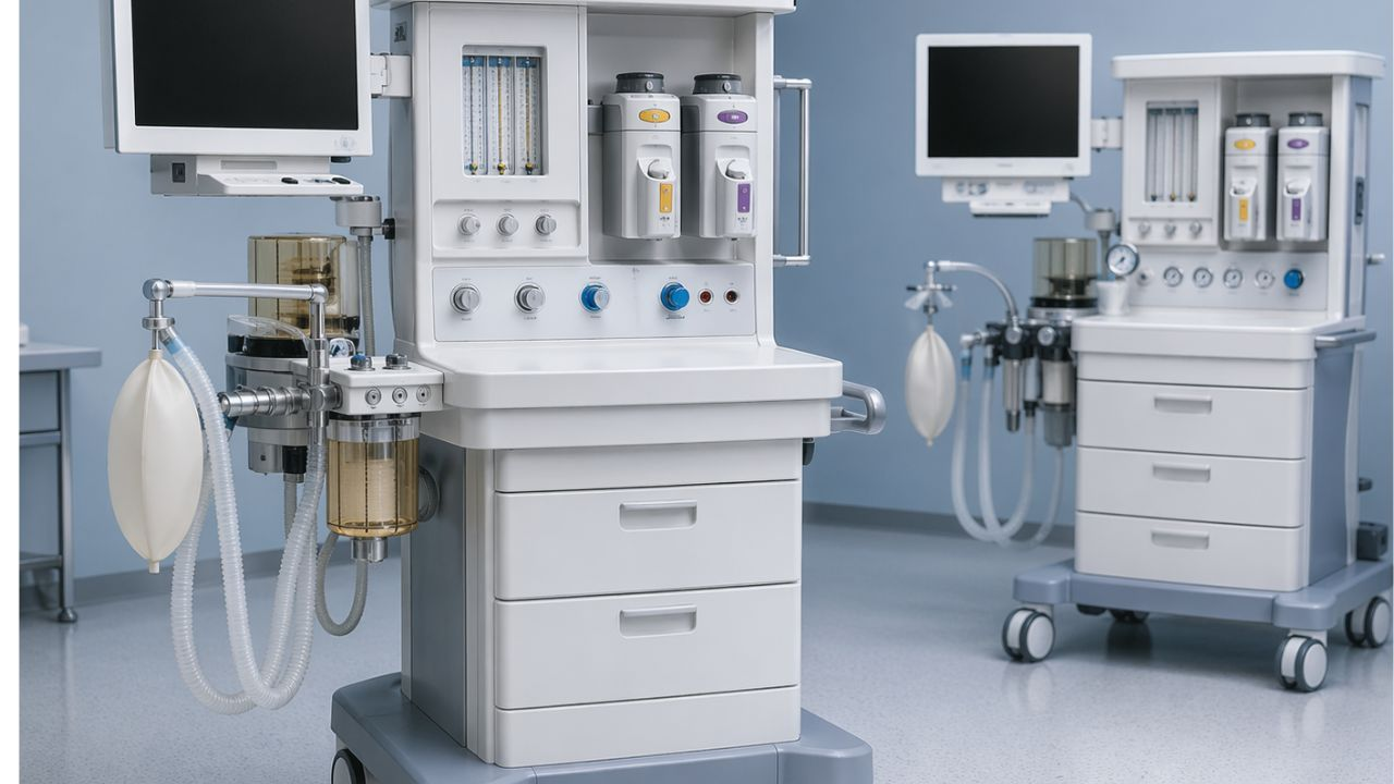
In a busy operating room, the line between a simple surgery and a serious emergency depends on the reliability of equipment and surgeons’ expertise. When a surgeon starts operating, they need tools that will make their job easy, which is exactly what you get with today’s anesthesia machines.
For the anesthesiologist, this machine is key. It’s the link between the patient being awake and them undergoing a pain-free, life-saving procedure. It lets them keep a close eye on the patient’s body functions while the surgeons do their thing.
Every successful surgery is backed by the confidence these machines give. Nowadays, companies make anesthesia machines with advanced breathing and monitoring features.
What is an Anesthesia Machine used for?
Basically, an anesthesia machine mixes oxygen and anesthetic gases to keep the patient asleep and doesn’t feel any pain. It also acts like a breathing machine.
The machine has fancy screens that show things like heart rate, blood pressure, etc. For the anesthesiologist, these numbers are like a way to talk to the patient’s body. If things change, the doctor tweaks the controls. It’s this continuous back-and-forth that keeps the patient in a safe, controlled state of unconsciousness.
What are the 4 types of Anesthesia?
When it comes to surgery and other medical procedures, four main types of anesthesia can be used. They are:
1. General Anesthesia
This type of anesthesia puts a patient into a controlled sleep. The anesthesiologist gives the patient strong medications that stop pain signals from getting to the brain. When the person wakes up, they won’t remember anything about the surgery.
2. Regional Anesthesia
The regional anesthesia numbs a larger part of your body, like the arm, the leg, or the whole lower half. They inject medication near a group of nerves to create a temporary “no feeling” zone for pain. However, an epidural, for example, is injected into the spinal canal and can also be used for childbirth.
With this, the patient can stay awake and chat with the doctor, but your lower body won’t feel a thing during the procedure.
3. Monitored Anesthesia
Often referred to as “twilight sedation,” the monitored anesthesia puts you in a sort of dream-like state. You’re not completely unconscious but relaxed and won’t notice any surgery taking place on your body.
This is mostly used for quick procedures. It’s a nice middle ground between being fully awake and completely asleep.
4. Local Anesthesia
The local anesthesia is the most basic type, used for small surgeries like cuts or biopsies. A numbing liquid (Betadine) is applied to the skin, in the mouth, or injected into the tissue. Local anesthesia is good for procedures that don’t take too long, usually under two hours, such as dental work or taking small tissue samples.
While it’s usually very safe, some people may have reactions like allergies or feel some tingling, burning, or swelling.
Top Anesthesia Machines
Selecting the most suitable anesthesia machine depends on factors such as the hospital’s budget, the clinical requirements, and the preferences of the anesthesiologists. Here are some of the top options available.
GE Healthcare’s Aisys CS2
The Aisys CS2 is a modern version of long-standing operating room machines. Its digital features ensure precise gas delivery and smooth integration with electronic health records.
- Advantage: It has a clean interface and enough ventilation. Also, the parts are easy to get.
- Disadvantage: Expensive due to the high-tech digital features.
- Popular because it’s familiar to those who trained on Ohmeda machines.
Dräger Anesthesia Machines
Dräger’s Perseus A500 is a sophisticated anesthesia machine known for its sleek design and automation. It adapts well to different patient sizes and includes automatic lung recruitment operations.
- Benefit: Saves space
- Advantages: Has a beautiful custom screen layout. Doesn’t make noise and does much of the work for you.
- Disadvantage: Requires software training.
- Popular due to the automation and advanced ventilation options.
Mindray: A9
Mindray used to be considered the “budget” alternative, but the A9 has transformed that notion. It’s not as expensive as the Aisys CS2; however, it serves well.
The benefit is that it serves as a “workhorse,” dependable, and user-friendly.
Key features include a large, easy-to-read touchscreen and an integrated “High-Flow Nasal Cannula” mode.
Advantages: Offers excellent value for the price. You get high-end features (such as improved ventilation).
Disadvantages: The build quality is acceptable, but not durable like previous GE or Dräger units. Some clinicians still need to familiarize themselves with the UI.
Why hospitals like it: For hospitals that desire high-end technology, it is still affordable.
IntelliSave AX700 from Philips
It makes perfect sense that Philips has an anesthesia machine that communicates easily with their monitor systems. The company is famous as a reliable brand for patient monitoring.
- Benefits: If your hospital is already a “Philips shop,” this makes more sense. The flow of data is incomparable.
- Key Features: It’s incredible how it integrates with Philips patient monitors. It lessens the “alarm fatigue” that so many medical professionals experience.
- The Advantages: Unified. Instead of feeling like a machine and a monitor pasted together, it works as a single, coherent system.
- Disadvantages: It is nearly overly integrated. The device may feel a little limiting if you don’t like the Philips ecosystem.
- Popular for: The data storage is the main reason people go for it. This device is perfect for anesthesiologists who enjoy having all their patients’ vital signs and breathing data in a synchronized stream.
Prima 465 from Penlon
The Prima 465 is designed to withstand the demands of a busy OR.
Important Features: It has a “ventilator with touch-screen control” but still has a very tactile, user-friendly appearance. It is renowned for being incredibly small.
Benefits: It’s a “back-to-basics” device that respects the anesthesiologist’s time. It is dependable, follows instructions, and doesn’t malfunction.
Advantages: It is very simple to maintain and clean. Its compact footprint is suitable in small operating rooms.
Disadvantages: Some of the most sophisticated AI-driven ventilation modes available on the high-end Dräger or GE versions are absent.
How to Buy Anesthesia Machines
If your hospital is planning to get a machine for anesthesia, here are tips to consider:
Evaluate the scope of your clinical work.
Knowing the “who” and “where” of your facility is essential before you make your choice. For a specialist dental surgery room, the equipment made for a high-volume trauma hospital is excessive.
- Patient profiles: Do you primarily treat adults, children, or newborns? Sensitive neonatal modes on certain equipment are vital for smaller patients.
- Complexity of Procedure: Are you doing simple outpatient operations or complex neurosurgeries that take hours? For the second, your clinic needs an advanced ventilation mode (such as SIMV or PRVC).
Give user experience and ergonomics top priority.
Usually, anesthesiologists stay close to this equipment for eight to ten hours during operation. So, the interface should be user-friendly.
- Intuitive Controls: How responsive is the touchscreen? Are the alarm systems easy to deactivate and read? You don’t want to be searching through submenus to change the oxygen flow in an emergency.
- Workspace Design: Does the device have enough shelves for charts and monitors? Are the drawers quiet and smooth? The anesthesiologist’s cognitive load is lessened by a simple workspace.
Assess Lifecycle Expenses and Reliability
You may want to evaluate the “Total Cost of Ownership.
- Maintenance Contracts: Find out whether local service specialists are nearby. How many hours will your operating room be offline while you wait for a part if the equipment breaks down?
- Consumables: Check the price of soda-lime canisters, O₂ cells, and proprietary sensors. Sometimes, a cheaper machine costs more because it needs expensive, brand-specific filters.
The Inspection Checklist
If you are buying a refurbished or pre-owned anesthesia machines, check the following:
Calibration History: Request the service logs. Has it undergone an annual professional calibration?
- Battery Backup: Examine the internal power supply. Make sure that the battery can last at least 30 to 60 minutes if the facility loses power.
- Software Updates: Know this: The safety precautions present in more recent software versions might not be present in older devices.
Involve the Final Users
The most important step is this one. Never buy an anesthetic machine without allowing your anesthesia team to “test drive” it.
Practical Demos: Ask suppliers to visit your establishment with a demonstration unit. Do a mock setup with your CRNA or lead anesthesiologist.
The “Feel” Factor: Find out from the clinicians whether the flow feels seamless. Is it simple to connect the breathing circuit? Do you think you can use this machine with a patient?
Reliability is essential in a high-stakes operating room. Make sure the unit has the necessary FDA, ISO, and CE certifications before deciding. These badges assure you that the device has met the international standard for patient monitoring, pressure control, and safety.
“Never concentrate only on the eye-catching touchscreen interface. Order the manual override features at all times. Make sure that your backup gas delivery and ventilation systems can function under high pressure.”
Future-Proofing on Anesthesia Machines
Nothing is more annoying than spending money on equipment that feels “old” as soon as it is installed. Future-proofing includes software upgradeability and modularity in addition to glossy screens.
Instead, look for machines that enable you to add new ventilation modes or sophisticated monitoring tools through software upgrades.
Do you think this machine can adapt if the care standards change in five years? Consider these:
Likability
Attempting to maintain patient stability while writing down numbers or manually transcribing data. It diverts your attention from the patient.
Your digital ecosystem is modern devices. Go for smooth interaction with your electronic medical records (EMR) or anesthesia information management system (AIMS). You want a computer that exports data automatically? Then get the machine connected to your EMR with ease.
Low-Flow Anesthesia
Low-flow anesthetics are becoming popular, and for good reason. In addition to lowering the quantity of costly anesthetic chemicals released into the atmosphere, it makes the patient’s breathing environment warmer, which may enhance their recuperation.
Read also: Best Surgical Tables in 2026 ( Uses, Features and Benefits
Wrapping Up
High-end anesthetic workstations are unquestionably important in today’s operating rooms. The standard in gas delivery and ventilation management is provided by devices such as the GE Aisys CS2 and the Dräger Perseus A500.
If you combine easy-to-use user interfaces with seamless patient monitoring, these machines reduce the cognitive load during critical procedures.
Medical teams put patient safety first by investing such dependable hardware. Meaning that with these instruments in a surgical suite, it changes the therapeutic experience.
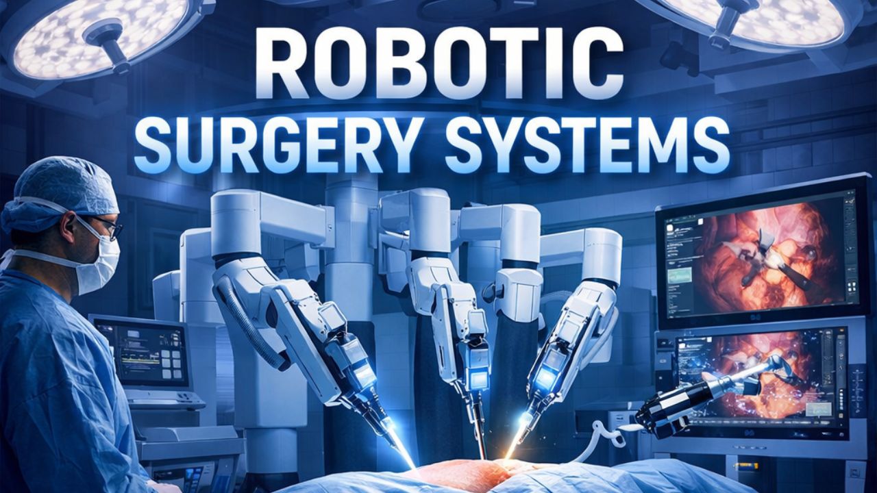
The phrase “robotic surgery” refers to a broad category of surgical platforms that help to facilitate, enhance, or automate tasks human surgeons perform. These platforms include teleoperating systems (controlled by a surgeon), telerobotic systems, and autonomous action (robotic surgery systems).
Some hospitals now center on robotic-assisted surgery. However, recent analysis highlights that questions are yet unanswered: What are the differences in clinical results between surgery performed with robotic assistance and surgery performed traditionally?
What are Robotic Surgery Systems?
With AI, tasks are performed faster, and the healthcare sector is not missing out on this automation. Machines now assist surgeons during operations. The robotic surgery system operates in this manner.
A mechanical arm, fitted with tiny tools, does the cutting and stitching. The doctor watches on a monitor and controls the robot with his/her hands. Basically, this setup provides more precision than hands alone. However, not every hospital uses these systems. Surgeons still make all key decisions.
One piece makes up part of what you see. Another section works behind that one. Then there is a third element that sits separately, but it connects closely to both.
A tiny lens brings sharp images into view during operations. This tool shapes how doctors see inside the body. The surgeon controls each tool. Every motion of the camera follows their hands. This is where precision begins.
With a robotic setup, surgeons can get accurate, smooth movement, and stronger command throughout the surgical operation. Through small cuts in the skin (minimally invasive), robotic surgery moves into the body. This method slips past thick layers of tissue gently.
There are little scars after the operation. In addition, it heals faster. Pain levels drop during recovery thanks to less damage inside. The risk of infection lowers because the openings are smaller. Also, the organs around the surgical area don’t get affected. More importantly, the probability of getting an infection after surgery is slim.
What are the Types of Robotic Surgery?
These machines assist urologists in treating prostate, kidney, and bladder issues. Also, gynecological use of this system involves removing the uterus or fibroid tissue. Even common operations such as fixing hernias or taking out gallbladders benefit from robotic surgery. Interestingly, patients don’t lose much blood during the surgery. To add, complications after surgery are rare, unlike traditional surgery.
Robotic Surgery Stystems By Specialty
Urologic Surgery
Surgeons turn to radical prostatectomy when treating prostate cancer. With robotic help, they take out the prostate but move carefully around nearby nerves. This care helps men keep control over urination after surgery. Sexual function stands a better chance, too.
With partial and radical nephrectomy, a robotic system allows careful removal of diseased parts and still protects working tissue. When treating cancer of the kidney, urologists rely on accuracy found through machine-assisted techniques.
Bladder surgery might take out just a section or the entire organ when dealing with cancer. This kind of operation usually leads to careful rebuilding that demands precision. Sometimes, what comes after depends on how much tissue is affected.
Gynecologic Surgery
- Hysterectomy: Surgical removal of the uterus, are used to treat conditions such as severe bleeding, endometriosis, or cancer. With robotic assistance, creates greater precision in tight anatomical areas.
- Myomectomy: Selective excision of uterine fibroids helps preserve the uterus. It involves smaller incisions compared to traditional open surgery.
- Oophorectomy or Ovarian Cystectomy: The removal of one or both ovaries or ovarian cysts, are used in the treatment of endometriosis or ovarian growths.
Gastrointestinal Surgery
- Cholecystectomy: The gallbladder is removed with robotic assistance, also enables a minimally invasive approach.
- Hernia Repair: Robotic technology have superior visualization, provide accurate mesh placement for both simple and complex abdominal wall hernias.
- Colectomy or Bowel Resection: Portions of the colon can be removed with enhanced precision, and the robotic system supports more controlled reconstruction of the intestinal tract.
Cardiothoracic Surgery
- Mitral Valve Repair: Heart valve repairs are performed through small incisions with robotic tools to prevent the need for an open-chest surgery.
- Cardiac Tissue Ablation: Abnormal heart tissue that causes arrhythmias is targeted and treated with robotic precision.
- Lung Resection and Tumor Removal: Pulmonary tumors or nodules are excised using robotic techniques that improve access to confined areas within the chest.
Head and Neck Surgery
Transoral Robotic Surgery (TORS): Tumors located in the throat, base of the tongue, or tonsils may need external cuts.
Orthopedic Surgery
Joint Replacement (Knee and Hip): Robotic-arm systems such as Mako use personalized 3D imaging to get accurate bone preparation, optimize soft tissue balance, and enhance implant positioning.
What are the 5 Commonly used Robotic Devices?
The top 5 robotic devices seen in many industries, such as manufacturing and medical, include articulated robots (assembly and welding robots), autonomous mobile robots (AMRs) (transportation robots), SCARA robots (fast pick-and-place robots), cobots (robots working alongside humans), and robotic surgical systems (precision surgery robots). Get the details below:
1. Articulated Robot (Industrial Robotic Arm)
This is the most common industrial robotic device. It has 3 to 6 rotating axes that resemble the human arm. Often used for welding, painting, material handling, and machine tending in automotive and manufacturing industries.
2. Autonomous Mobile Robots (AMRs) & AGVs
Robots such as autonomous mobile robots (AMRs) and automated guided vehicles (AGVs) use sensors, cameras & GPS to move parts or finished goods. They are used in warehouses, factories and hospitals. Amazon warehouse robots and the TUG are two popular examples.
3. SCARA Robots
Highly accurate & speedy robots used for precise vertical assembly movements. SCARA robots (as they use 4 axes) are used widely in the electronics industry as well as the car manufacturing industry for tasks including component insertion, packing & assembly.
4. Collaborative Robots (cobots)
Unlike industrial robots, cobots work alongside workers in a factory. They can be quickly programmed for different uses and include tasks such as kitting, machine tending, and packing.
5. Robotic Surgical Systems (medical robots)
These systems are designed for less invasive surgery and allow surgeons to perform complex procedures using robotic arms that has better control and in 3D capacity. For instance, the da Vinci Surgical System, orthopedic robots (e.g., Mako) and robots for sterilization purposes (e.g., Xenex).
The Top Robotic Surgery Systems
Intuitive Surgical: da Vinci Systems (Xi, X, SP, 5)
The industry standard for soft-tissue surgery, for urology, gynecology, and general surgery uses the da Vinci robotic systems. It features high-definition 3D imaging, wristed instruments (EndoWrist), and the new da Vinci 5, that have a force feedback and advanced imaging.
Specialty Platforms: Ion (endoluminal robotic bronchoscopy for lung biopsy).
Stryker: Mako SmartRobotics
Focus: Orthopedic joint replacement (knee and hip).
Details: It uses pre-operative CT scans to create a 3D model, allows surgeons to customize implants and guide robotic arms for precise bone cuts.
Medtronic: Hugo RAS System
It’s for soft-tissue procedures: Medtronic has a modular, open-console design that enables hospitals to use independent robotic arms. It enhances flexibility and affordability.
Specialty Platforms: Mazor (robotic guidance for spine surgery).
CMR Surgical: Versius
Focus: Minimally invasive, versatile surgery.
It’s portable, and designed to fit into existing surgical workflows easily. It is increasingly used in Europe, Australia and India for various laparoscopy procedures.
Asensus Surgical: Senhance System
- Focus: Laparoscopic digital surgery.
- Details: Emphasizes haptic feedback (touch sensation) and eye-tracking camera control, with a high precision.
Globus Medical: ExcelsiusGPS
- Focus: Spine and cranial surgery.
- Details: Provides real-time imaging and robotics-guided precision for spinal fusion and other neurosurgical process.
Zimmer Biomet: ROSA Robotics System
- Focus: Orthopedic surgery.
- Details: Specialized in robot-assisted knee and hip replacements with advanced data analytics for precise positioning.
- Focus: Robotic bronchoscopy.
- Details: Used for early-stage lung cancer diagnosis.
Robotic Surgery Systems’ Growing Effect on Modern Healthcare
Because of smaller incisions used during robotic-assisted surgery, the patient will experience fewer blood losses (an average of 50.5% decrease in loss of blood), a reduced need for transfusions, a shortened recuperation period, and small instances of postoperative infections.
Better control for the surgeon: Robotic systems enable the use of 3D and visualization, which offer a clear and high-resolution view of the surgical site. The robotic arms offer greater dexterity and tremble cancellation and enable the performance of tasks far greater than those performed by human hands.
Growth of new specialties using this technology: While the technology was first widely implemented for prostatectomies within urology, other specialties have adopted it for:
- Gynecology for hysterectomies and endometriosis surgery
- General surgery for hernia repairs, gallbladders, and bariatric surgery procedures
- Orthopedics for joint replacements
- Cardiothoracic surgery for heart valve repair procedures.
Robotic-Assisted Surgery’s System Improvement
Use of AI and Modern Technology
Robotic surgery is getting popular by the day, as leading surgical platforms now use artificial intelligence (AI) to improve decision-making, analytics, and the possibility of personalized medicine. Haptic technology has also become a growing implementation in robotic systems, as it allows surgeons to feel tissue resistance.
Benefits and Challenges of Robotic Surgery
The advantages of robotic surgery include better accuracy, less blood loss, a lower infection rate, and quicker recovery for the patients. Improved visualization and comfortable ergonomics benefit the surgical teams.
Cost, the difficult learning curve for surgeons, and the possibility of technical problems and an increase in operating time are the main disadvantages.
Advantages for the Patients
Increased Speed of Recovery & Less Pain: Patients usually spend less time in the hospital and experience less pain and have a more rapid return to normal life than is typical with conventional surgery.
Decreased Complications: With smaller incisions, there is less risk of infection and a decreased rate of blood loss and clotting.
Increased Accuracy: The ability for the system to make high-precision movements necessary in complicated, meticulous surgery like that done in urology, gynecology, and cardiology.
Improved results in difficult cases: For instance, in robotic prostatectomies and colorectal surgery.
Advantages for the medical team (surgeons and staff)
The 3-D, high-definition, and magnified view available through the system provides the surgeon with the best view possible of the operative field.
The dexterity of the robotic arms is superior to the range of motion and filter for tremors possible with conventional laparoscopy.
The surgeon’s access to improved ergonomics through the seated console position reduces fatigue in the neck, back, and shoulders.
Problems Patients Face
- Cost: Costs can be higher than that of traditional procedures. So, hospital will charge more. Although, it is quicker.
- Possibility of failure: There are risks that an instrument or the robot will malfunction, and that will require it is converted to open surgery.
- Not Suitable for All: Robotic surgery isn’t appropriate for all patients. Discuss with our doctor before using one.
Problems the Medical Team Faces
- Learning Curve: Large amounts of training and simulation time are needed to master the robot system.
- Expensive Investment & Maintenance: Most well-funded hospitals can only afford the cost of the initial acquisition of the robots, including their maintenance.
- No haptic feedback: Surgeons can no longer ‘feel’ with their hands; all surgical feedback must be taken from the camera system.
- Longer setup time: The setup time of the robotic system may increase more time.
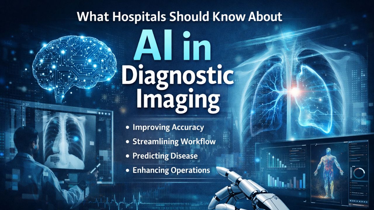
Read also: AI in Diagnostic Imaging: What Hospitals Should Know Before Adopting It
Wrapping Up
A robotic surgery system is identified as a surgical robot for minimally invasive surgery. There are two categories: independent robotic surgery systems, in which the entire surgical process is carried out robotically, and assisted laparoscopic surgery systems, where robotic-assisted surgery is used in conjunction with laparoscopy for specific aspects of the procedure.
However, hospital that want to set this should consider the need, capital and and potential return. The best solution must be viewed with a long-term perspective, considering both the investment and benefits.
The questions that addresses robotic surgery systems are: Will the clinical benefits make it a worthwhile investment for a hospital to improve the quality of its service, attract cutting-edge surgeons, and market itself to a wider customer base? and Will the anticipated returns excite an investor with a higher capacity to invest?
We consider investment return based on clinical benefits, effects on the patient, and learning. So, the factors that support or affect the hospital’s decision depend on the hospital’s mission, investment model, and age of patients.
Diagnostic & Hospital Equipments
AI in Diagnostic Imaging: What Hospitals Should Know Before Adopting It
If someone told you ten years ago that machines will do the work of a doctor or radiographer, you’d probably laugh, right? But today, that’s our reality. AI in diagnostic imaging isn’t some sci-fi fantasy anymore; it’s quickly becoming part of regular medical practice. However, adopting an AI-powered system comes with challenges.
There’s the tech side, integrating it with your existing workflows, training staff, and ensuring that the analyses are accurate, and then there’s the human side, which can be even more complicated, no matter how advanced the technology is.
What is AI in Diagnostic Imaging?
When I mention AI in diagnostic imaging, I’m referring to the software and systems that help interpret medical images such as CT scans, MRIs, X-rays, and ultrasounds. This technology leverages machine learning and deep learning to sift through pixels, detect patterns, and identify areas that need attention. Imagine computer vision taken to the next level.
Some systems evaluate images before a radiologist even takes a look; others provide an additional perspective. These tools can point out things a human might overlook, speed up results, and in some research, and match the diagnostic accuracy of seasoned specialists. That’s a significant development. However, hospitals can’t buy a system and consider their work done.
Why AI Is Important in Imaging
The impact of AI on medical imaging cannot be overemphasized. Consider the usual process: a patient gets scanned; the radiologist looks at the images, and then writes a report. This method can be slow. However, introduce AI into this sequence, and everything changes:
- Information flows more quickly.
- Radiologists receive insights within context.
- AI detects subtle patterns that might escape human eyes.
- Consistency becomes better, especially in busy environments.
So, the whole imaging process speeds up and becomes more efficient.
Sometimes, AI can almost match the skills of expert radiologists in specific tasks. In mammography, for instance, these tools have successfully identified cancerous growths with precision that rivals human-only examinations.
Interestingly, this technological advancement has led to a significant improvement in early detection rates. Moreover, AI’s role continues to expand as it becomes more integrated into diagnostic processes. Technology is not just supporting but also enhancing the capabilities of medical professionals.
It doesn’t just spot disease early; it improves patients’ well-being. Also, it’s not just in regular radiology anymore. You’ll find these applications in analyzing pathology slides and cardiac images, among others. They even use foveated models that focus on important areas, similar to how our eyes naturally work.
Areas Where AI Brings Benefits
When hospitals start using AI-driven imaging systems, they can enhance how they care for patients, boost the confidence of their medical staff, and make departments run more smoothly. Here’s why these systems are so valuable:
Better Diagnoses
AI can look through thousands or even millions of scans with labels, spotting patterns and tiny issues that might escape human notice. Some systems zoom in on areas likely to have problems, similar to how our eyes focus on important details.
This tech doesn’t replace radiologists; it gives them a helping hand. As it offers extra insights, AI makes readings more accurate, cuts down on overlooked findings, and helps catch things early, like lung nodules or beginning tumors. Doctors get an extra pair of eyes from the system, which makes them trust their analysis.
Improve Workflow Efficiency
Imaging backlogs can delay patient care and put pressure on radiology teams. AI steps in to sort scans, highlight urgent or high-risk cases for quick review, and manage standard ones. Automating repetitive tasks like labeling, taking measurements, and preparing initial reports, cuts down on workload and errors.
When departments include AI to support their workflow, they see faster processing times, which can lead to a smoother imaging process too.
Predictive Insights
Some AI tools don’t just look at images on their own. They mix scan data with a patient’s past records to spot diseases forming before any symptoms appear. Take cardiac imaging, for instance. AI finds tiny changes in heart tissue that might lead to problems later. These predictive skills add extra depth to the analysis and assist care teams in making smarter choices that could boost patient health.
Operational Support
AI is transforming how imaging departments run. It takes care of tasks like scheduling and preprocessing images, ensures quality, and keeps an eye on equipment. Some systems even keep track of scanner performance as it happens, spot calibration problems, and catch data issues before they reach the doctors.
Meanwhile, other systems make sure that images look the same no matter who’s on shift. As a result, radiologists and technicians get more time to concentrate on interpreting results and make clinical decisions. This shift boosts both efficiency and the quality of patient care.
Challenges of AI in Diagnostic Imaging
Implementing AI in imaging is not about installing the software. There are challenges hospitals need to understand before they set any AI-powered system. These are not impossible, but they require planning, and cooperation from IT, clinical teams, and administration. Some of the limitations are:
Integration Isn’t Easy
Bringing new technology into hospitals is not a walk in the park. Hospitals rely on systems like Picture Archiving and Communication Systems (PACS) and Electronic Health Records (EHR). Adding an AI system into this mix is not as easy as just plugging it in.
Strong technical infrastructure, solid network security, and well-designed workflows are essential for safeguarding patient information. Even moving images for analysis can lead to compliance and privacy challenges.
Building these systems calls for more than just IT experts; you need people that understand both medical tech and clinical demands.
Trust Issues
AI learns from the data it encounters. So, if a system gets trained on data that doesn’t match your patients, it might not perform well. For instance, a model focused mainly on adult scans might find pediatric imaging challenging.
Similarly, an AI developed in one country could misinterpret images from another region’s patients. This poses a genuine issue since clinicians depend on accuracy.
If the AI frequently overlooks certain cases or wrongly flags normal scans, health providers won’t trust the system. Hospitals must assess AI tools thoroughly and ensure their training data aligns with their patient demographics.
The Black Box Problem
Many AI models, particularly deep learning systems, are often compared to black boxes. They produce results without offering clear explanations for their decisions. A radiologist might see a lesion marked by the system but lack insight into the reasoning behind it.
This lack of transparency can make clinicians wary of trusting AI. Although explainable AI is on the rise, decisions without clear reasoning can lead to even the most accurate systems being overlooked because doctors need assurance that the analysis they receive is dependable.
Regulatory and Ethical Layers
In some countries, regulations are clearly defined; in others, not so much. Hospitals require a compliance plan that addresses data privacy, liability, and ethical use. Think about it: if an AI system fails to diagnose correctly, who takes the blame? And how should you record analyses done with AI in patient files?
These aren’t just hypothetical questions; they play a significant role in everyday hospital operations and legal matters. Addressing regulatory and ethical issues is vital for technical integration.
What Hospitals Should Consider Before Adopting AI in Diagnostic Imaging
Health Facilities should take note of the following:
1. Have a Goal
AI systems differ; some focus on stroke detection, while others specialize in mammography or lung scans. A multipurpose software may seem convenient, but it’s not always the best. Before you invest, consider the problem you want to address.
- Do you need to speed up critical case detection?
- Or do you want accurate readings?
The response will guide your facility to make the right decision.
2. Focus on What Matters
Consider this: Are scan reading times improving? Do radiologists feel less stressed? Do patients receive their results quickly now? It’s important to establish clear goals for workflow, staff morale, and clinical outcomes. This way, you can see if the AI system is truly effective.
3. Start Small
Implementing AI on all imaging devices might cause mistakes among the staff. Instead, focus on one area first. Choose a scanner, department, or case type for a trial run. Use it for several weeks to evaluate its performance. Adjust settings based on observations and collect feedback from the team during this time.
Integrating AI gradually, allow it to blend smoothly into the workflow without interfering with everyday tasks.
4. Educate Your Team
Even the best AI system won’t help if no one knows how it works. Your team needs to understand how to interpret AI results and what the confidence levels mean. They should also know when to trust alerts or question them. Learning is not a one-time thing. As the AI system evolves, your team should get updates so they stay on track.
Understanding the Costs of Adopting AI in Imaging
Bringing AI into your imaging department goes beyond just paying for a license and hitting “install.” There’s more to it, and hospitals need to plan wisely. The expenses can be divided into key categories:
Integration and IT support: AI must work seamlessly with existing systems like PACS and EHRs. This often means additional IT tasks, hiring specialized staff, and ensuring ongoing tech support.
Data governance and security infrastructure: Patient data is sensitive. Strong protections are essential to maintain privacy, follow regulations, and prevent breaches. New policies are frequently necessary for AI systems.
This journey requires thoughtful preparation across various areas. Secure storage and monitoring tools are essential. Staff needs training to grasp system usage, interpret AI results, and understand its limits.
Without proper education, even advanced AI tools might not be fully implemented. Hospitals must ensure the system works for their patients before relying on AI; this involves pilot programs, internal studies, and continuous reviews.
Don’t just consider the initial cost. Adopting AI is an investment that enhances patient care and departmental efficiency but comes with responsibilities and expenses.
Final Thoughts on AI in Diagnostic Imaging
AI is shaking up medical imaging, and it’s happening faster than many predicted. It helps doctors detect disease faster speeds up report processes, and enhances care precision. However, AI has its limitations; it won’t manage your department for you. Before integrating AI, hospitals should consider some important questions:
- What do we need from this system?
- How does it assist doctors in performing their tasks?
- What metrics will show if it’s effective?
- Are we ready to train staff, change workflows, and update the software when necessary?
Tackle these questions first. If satisfied, go ahead and integrate AI in your hospital or clinic.
-
 Uncategorized4 months ago
6 Best Heatmap Plugins – I Test, Review and Compare
-
 AI in Health care3 months ago
Remote Patient Monitoring in Modern Healthcare
-
 Diagnostic & Hospital Equipments4 months ago
What Are Medical Devices? Types and Uses
-

 Review and guides3 months ago
CPAP Machines Buying Guide: The Best Models Doctors Don’t Recommend
-

 AI in Health care1 month ago
Digital X-ray vs. Film X-ray: Which is better?
-
 AI in Health care3 months ago
AI in Medical Imaging: What is Coming Next
-
 Diagnostic & Hospital Equipments1 month ago
DEXA Bone Density Scanning: What You Need to Know About Bone Health
-
 Diagnostic & Hospital Equipments4 weeks ago
PET Scan Machines: Understanding the Technology, and Usage